
Corynebacterium diphtheriae (Diphtheria)
Authors: Sabrina Weiss and Androulla Efstratiou
Author, Second Edition: Christine C. Chiou, M.D.
MICROBIOLOGY
Corynebacterium diphtheriae is the leading causing agent of diphtheria. It is a non-motile, non-encapsulated, non-sporulating gram-positive rod-shaped bacterium with a high GC-content and occurs in four biovars: gravis, mitis, intermedius, and belfanti, based on colonial morphology and biochemical profiles. Microscopically, corynebacteria have club-shaped ends and display a characteristic v-shaped morphology. In addition to C. diphtheriae, two other corynebacteria species can produce diphtheria toxin and thus also cause diphtheria: C. ulcerans and very rarely C. pseudotuberculosis. Both are zoonotic pathogens that can have the ability to produce diphtheria toxin (29, 52).
The ability to cause diphtheria depends on the organism’s ability to produce diphtheria toxin. The respective tox gene is encoded on the genome of a lysogenic β –phage, but its expression is regulated by an iron-dependent chromosomal repressor (56). The name diphtheria is derived from the greek word ‘diphthera’ meaning ‘hide, leather’, and refers to the characteristic membrane that is provoked by the respiratory disease caused by a toxigenic strain.
Non-toxigenic strains can also cause disease; respiratory symptoms are milder and do not present with a pseudomembrane. The malicious characteristic of the non-toxigenic strains is that they can be invasive causing bacteremia, endocarditis, abscesses, and bone and joint infections (24). The majority of these strains completely lack the tox gene, but non-toxigenic tox-bearing (NTTB) isolates that do not express the toxin have also been described (56). In contrast to toxin-induced disease, infections with non-toxigenic strains are not preventable by vaccination.
EPIDEMIOLOGY
C. diphtheriae is transmitted between humans via droplets, secretions or direct contact. In the United States and Europe, infections with toxigenic C. diphtheriae have become uncommon and cases are mostly imported from endemic areas. In non-endemic countries the circulation of toxigenic and non-toxigenic strains as well as the correlation between colonization and vaccination is still not completely understood (56). Widespread vaccination campaigns have made diphtheria an uncommon disease in some parts of the world, but it is still endemic in South America, Eastern Europe, Southeast Asia, and Africa (18).
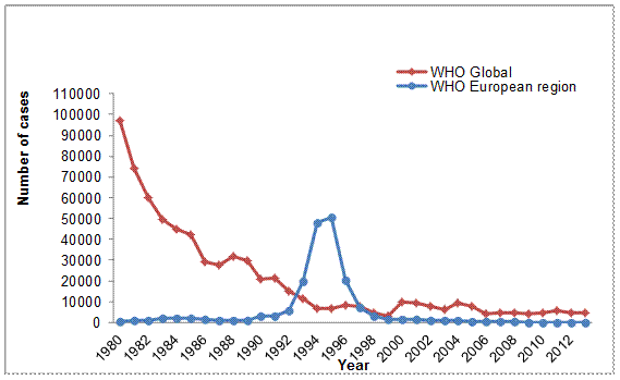
Diphtheria was previously known as a childhood disease and owing to comprehensive routine vaccination programs, coverage is now mostly high in children within developed countries. In countries where vaccine coverage is low, circulation of toxigenic C. diphtheriae continues. In 2013, 4,680 diphtheria cases globally were reported to WHO (54), 80% of which occurred in just two countries – India and Indonesia. The number of cases reported from countries mainly from within the South East Asia Region continues to rise and there is an urgent need to address this (Figure 1). Current priorities include improving surveillance systems, laboratory diagnostics and typing methods as well as identification of population immunity gaps and determinants of poor vaccine coverage. An estimated 2500 deaths from C. diphtheriae occurred in 2013. Global estimated third dose diphtheria/tetanus/polio vaccine (DTP3) coverage in 2013 was 84%, but only 30% of countries reached the target of >=80% DTP3 coverage in all districts (50). Control of diphtheria seems to rely on good coverage in childhood, as seen in the example ofEngland and Wales where data from 1914-2012 showed the dramatic disappearance of diphtheria cases and deaths, and continued the low numbers of cases despite the fact that the proportion of adults with full protective immunity (antitoxin ->= 0.1 IU/ml) is quite low (less than 30% in the older age groups) (38).
Diphtheria usually appears ‘periodically’. The last huge epidemic wave occurred during the 1990s in Russia and the States of the former Soviet Union, and was characterized by a large proportion of adult/ adolescence cases. This was attributed to a lack of vaccination and/ or insufficient booster vaccinations in these age groups (8, 12, 18). Epidemics are still persisting in areas such as South East Asia and Africa where several thousand cases are reported annually, the numbers are underestimated due to the lack of surveillance infrastructure within these countries. This is currently being addressed for areas within South East Asia by WHO.
Despite the rarity of C. diphtheriae infections, disease incidence caused by C. ulcerans has been increasing in industrialized countries (18, 29). C. ulcerans is a zoonotic pathogen, most often found in pets and domestic animals and probably not transmitted between humans (29). Occasionally, in can be spread by contaminated food, e.g. milk (56).
Owing to a lack of robust surveillance or diagnostics in many countries the prevalence of C. ulcerans is not yet well understood and warrants investigation. However, this is hampered by the inconsistency of the case definitions for diphtheria. Currently two different case definitions are applied in public health settings across the European region: the 2008 EU case definition, which considers disease caused by C. diphtheriae, C. ulcerans and C. pseudotuberculosis, and the 1994 WHO case definition, which only considers classical respiratory diphtheria cases caused byC. diphtheriae (‘epidemic diphtheria’). Both definitions are detailed in Table 1 and differences discussed below. In addition, it is standard practice that case definitions may be modified during specific incidents or outbreaks. The Commission Decision 2008/426/EC lays down case definitions for reporting communicable diseases in EU.
Neither of the case definitions includes non-toxigenic organisms, which do not require any public health action (antibiotic treatment only). A subset of non-toxigenic C. diphtheriae strains are known to carry the tox gene [non-toxigenic toxin-gene bearing (NTTB) strains (52). During and after the diphtheria epidemic in the former Soviet Union, the circulation of NTTBs was observed (34), and two NTTBs were detected in Lithuania in a recent screening study (47), but in general the prevalence of NTTB strains is ill-defined. NTTB strains are often isolated alongside other organisms (4, 31, 40). Although the potential of phage conversion to transform NTTBs into toxigenic strains is considered low and rarely observed in vivo, circulation of NTTBs may act as a repository for tox gene sequences and therefore pose a risk for disease. However, the public health relevance of these strains is yet to be examined and further studies are needed in order to assess the true burden of NTTB strains in Europe and globally.
![]()
CLINICAL MANIFESTATIONS
The two major sites of infection are the respiratory mucosa and skin, rarely extra-respiratory mucosal sites, e.g. eye, ear, or genitals can also be affected (8, 56).
Respiratory diphtheria
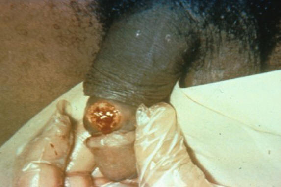
The incubation time of respiratory diphtheria is 2-5 days and initial symptoms include sore throat, malaise, and low-grade fever. The characteristic clinical presentation is the presence of a grayish-white, fibrinous and firmly adherent pseudomembrane (Figure 2) that forms within the first few days and spans over the tonsils, the pharynx, or the larynx. Laryngeal diphtheria is characterized by gradually increasing hoarseness and stridor and most commonly occurs as an extension of pharyngeal involvement in children. Nasal diphtheria, usually mild and chronic, is marked by uni- or bilateral nasal discharge, which is initially clear and later becomes bloody (3, 26). With disease progression, the membrane may become necrotic and exhibit green and black patches. Attempts to dislodge it usually result in bleeding. Extension of the membrane correlates with disease severity and suffocation following accidental aspiration is a common source of death. Marked swellings of cervical lymph nodes (lymphadenitis) and swelling of the surrounding soft-tissue give rise to the characteristic “bull-neck” appearance. If untreated, the disease usually lasts for 1-2 weeks and the case fatality rate is about 10% (24).
Most common complications are myocarditis and neuritis. These are attributable to absorption of diphtheria toxin at the site of primary infection and can last for months. Myocarditis presents as abnormal cardiac rhythms that can lead to heart failure and subsequently death (8). Neurological symptoms occur in 75% of patients with severe disease and can range from mild temporary paralysis of motor nerves to total paralysis (24). Paralysis of the diaphragm may result in secondary pneumonia and respiratory failure (8).
Respiratory symptoms can also be caused by non-toxigenic corynebacterium strains, but are usually milder and without membrane formation.
The management of a patient with suspected diphtheria includes:
· Administration of diphtheria antitoxin as soon as possible after testing for hypersensitivity to horse serum; early administration of appropriate antitoxin is critical for survival;
· Confirmation of the diagnosis through appropriate bacterial cultures;
· Administration of antibiotics; and
· Appropriate supportive care including special attention to maintaining an adequate airway in the presence of laryngeal or extensive pharyngeal membranes and to careful monitoring for cardiac rhythm disturbances or other manifestations of myocarditis.
Cutaneous diphtheria
Cutaneous diphtheria can be caused by either toxigenic C. diphtheriae or C. ulcerans and commonly occurs on exposed limbs, particularly the legs. The lesions start as vesicles and quickly form small, clearly demarcated and sometimes multiple, ulcers that may be difficult to distinguish from impetigo (Figure 3). The classic description of diphtheritic lesions is that they are usually covered with an eschar, a hard bluish-grey membrane that is slightly raised. They usually present as as non-healing ulcers that may be difficult to distinguish from other chronic bacterial infections (37). Cutaneous diphtheria is often associated with concurrent infections caused by Staphylococcus aureus or group A streptococci (56).
LABORATORY DIAGNOSIS
The main role of the laboratory in the diagnosis of diphtheria is to provide simple, rapid and reliable methods to assist clinicians in confirming a clinical diagnosis. In advanced cases a clinical diagnosis of diphtheria normally precedes the microbiologic diagnosis. However, it is often difficult to diagnose diphtheria on clinical grounds as the disease may be confused with other infections, such as severe streptococcal tonsillitis, glandular fever or Vincent’s angina. The laboratory may also aid the clinician by eliminating suspected cases or contacts of diphtheria from further investigation, thus avoiding unnecessary treatment or control measures such as isolation. Guidelines for the laboratory diagnosis of diphtheria have been published (14). These are currently being revised to include novel and up to date methods. Laboratory diagnosis usually relies upon the cultivation of bacteria from clinical specimens. Throat swabs (preferably taken from the membrane) or swabs from wounds or lesions are cultured onto blood and tellurite agar. Tellurite inhibits growth of some normal flora and allows the Corynebacterium sp. (and also other pathogens such as yeast and staphylococci) to grow as black or grey colonies. Corynebacteria are quite characteristic on this medium as small black colonies that require further confirmatory tests. Commercial kits (API®Coryne, API bioMerieux) allow for identification of the strain and biotype. More modern methods are being used increasingly in many laboratories such as MALDI-TOF MS (matrix assisted laser desorption/ionization time of flight mass spectrometry which is a new technology for species identification based upon the protein composition of microbial cells. The method allows rapid and reliable identification of clinically relevant and potentially toxigenic corynebacteria provided that a quality-controlled database of reference spectra is available (30). However, the absence of pyrazinamidase, or the presence of cystinase activity will identify potentially toxigenic species (C. diphtheriae, C. ulcerans, C. pseudotuberculosis), but the gold standard for detection of toxigenicity is the Elek immunoprecipitation test which indicates presence of a biologically active protein (17). The most important test in the microbiologic diagnosis of diphtheria is therefore, the detection of toxigenicity. Currently, the only in vitro phenotypic method readily available is the Elek immunoprecipitation test and is only undertaken by specialized reference laboratories. The modified Elek test which simplifies the toxigenicity testing of corynebacteria is recommended; a result is available in 16 to 24 hours (17). Real time PCR assays have also been described for detection of the toxin gene and are being used in many specialized reference laboratories (20, 36). However, any PCR positives must be confirmed by the phenotypic Elek test due to the possible although rare occurrence of NTTB strains (15).
![]()
PATHOGENESIS
The primary sites of infection for pathogenic cornyebacteria are mucous membranes where they induce formation of the characteristic pseudomembrane. Their pathogenesis largely depends upon toxin production. The toxin acts by inhibiting cellular protein synthesis by ribosylation of adenosine diphosphate leading to inactivation of elongation factor 2 (24, 32).
Diphtheria toxin is a very potent exotoxin consisting of 353 amino acids. The lethal dose for susceptible individuals is approximately 0.1 µg/kg body weight (55). Strains can have more than one tox gene inserted, which directly correlates with their toxigenicity (24). While the tox gene is encoded on the β –phage DNA, its expression is regulated by the chromosomal diphtheria toxin repressor DtxR. The repressor is regulated by iron supply in a way that a lack of iron leads to inactivation of the repressor protein, resulting in toxin production (27).
Pathogenicity of non-toxigenic strains mostly depends on their ability to become invasive and cause bacteremia. Whether and how NTTB strains differ in pathogenesis is not known. They carry the tox gene, but cannot express the toxin mostly due to genomic changes such as insertions or deletions resulting in frame shift within the open reading frame (56).
SUSCEPTIBILITY IN VITRO AND IN VIVO
In vitro, C. diphtheriae isolates are generally highly susceptible to erythromycin as well as to the newer macrolides (Table 2) (2, 21, 22, 57). Resistance to erythromycin has been reported in Canada and the United States. In Canada, nontoxigenic C. diphtheriae biovar mitis from two patients with chronic superficial skin lesions who had previously been treated with erythromycin and lincomycin were found to be highly resistant to both antimicrobials, although both remained fully sensitive to penicillin G (28). During the diphtheria outbreak in Seattle in the1970s, erythromycin was used extensively for treatment. Nontoxigenic C. diphtheriae biovar mitis isolates from patients with cutaneous diphtheria were found to have inducible resistance to both erythromycin and clindamycin when grown in the presence of subinhibitory concentrations of erythromycin (10). This resistance was plasmid mediated (42), and homology was demonstrated between this plasmid and plasmids found in skin coryneforms, suggesting that it may have originated in skin flora (43). After 1978, use of erythromycin for treatment of skin lesions among at risk populations was no longer recommended, and the frequency of erythromycin resistance of clinical isolates of C. diphtheriae decreased dramatically (25). Ciprofloxacin and other fluoroquinolones have demonstrated excellent in vitro activity against C. diphtheriae, but no clinical data are available on experience with these agents in the treatment of diphtheria. High level resistance to rifampin has been demonstrated in biotype gravis strains recovered from patients in Estonia, but the treatment history of the patients was unknown (33). Tetracycline resistance has been reported in Indonesia, where tetracycline was often used for empiric treatment of infections (41). Ampicillin tolerance of C. diphtheriae biotype mitis strains from patients with endocarditis has been reported (13).
![]()
ANTIMICROBIAL THERAPY
Antibiotic treatment is necessary to eliminate the organism and prevent spread; it is not a substitute for antitoxin treatment. All specimens should be collected before antibiotic treatment is started. The empirical choices of antibiotics are macrolides (erythromycin, azithromycin or clarithromycin) or benzylpenicillin, all of which are usually active against C. diphtheriae and C. ulcerans (49). If erythromycin cannot be tolerated (because of gastrointestinal side-effects) an alternative macrolide such as azithromycin or clarithromycin should be given. Parental benzylpenicillin or erythromycin should be used until the patient can swallow comfortably, then erythromycin or penicillin V should be continued.
The recommended dose regimens are as follows:
A. Penicillin, preferably intramuscular procaine penicillin G (25 000 to 50 000 units/[kg/d] for children and 1.2 million units/d for adults, in two divided doses) or parenteral erythromycin (40 – 50 mg/[kg/d], with a maximum of 2 g/d until the patient can swallow comfortably, at which point erythromycin in four divided doses or oral penicillin V (125 – 250 mg four times daily) may be substituted.
B. Antibiotic treatment should be continued for 14 days.
C. Although rarely encountered, resistance of C. diphtheriae strains towards commonly used antibiotics including erythromycin have been documented (42), knowledge of antimicrobial susceptibility patterns for strains recovered in individual locales is therefore warranted.
Respiratory Diphtheria
Antibiotics and antitoxin are both essential for treatment of respiratory diphtheria. Antitoxin should be given upon clinical diagnosis without waiting for laboratory confirmation (see section IV, adjunctive therapy).
The antibiotics of choice are macrolides. Erythromycin, orally or by injection (40 mg/kg/day; maximum 2 g/day), or procaine penicillin G daily, intramuscularly (300,000 U/day if 10 kg or less body weight, and 600,000 U/day > 10 kg body weight) should be given until the patient can swallow comfortably (3). At that point, treatment may be substituted by four doses of oral erythromycin or penicillin V (125-250 mg/day) for a minimum of 14 days total (3). All drugs show in vitro activity against C. diphtheria and C. ulcerans (16).
During convalescence all patients should receive a first or booster vaccination, since infection may not induce protective immunity (55).
Cutaneous Diphtheria
Treatment of cutaneous diphtheria includes thorough cleaning of the lesion with soap and water (3). Patients with cutaneous diphtheria should receive the same antibiotic treatment like patients with respiratory disease. However, administration of antitoxin is only indicated for patients with sufficiently large ulcers (i.e. more than 2cm2) or if they are membranous (37).
Special Infections
Myocarditis and Neuropathy
Myocarditis and neuropathy are the most common complications that result from excessive toxin absorption. Antitoxin must be given and conditions be treated symptomatically.
Endocarditis
Endocarditis due to C. diphtheriae has commonly been treated with a β-lactam antibiotic in conjunction with an aminoglycoside, but has also been successfully treated with β-lactam antibiotic alone. Concerns have been raised whether the benefits out-weight the risk of toxicity upon prolonged use of aminoglycosides and it has been proposed that monotherapy could be favored depending on patient-specific factors like age, preexisting renal impairment, or ototoxicity (35).
Endocarditis cases are mostly associated with non-toxigenic C. diphtheriae so antitoxin is not required for treatment.
Zoonotic infections
Treatment of infected companion animals to eliminate subclinical carriage of toxigenic C. ulcerans may be thought appropriate in some cases (e.g. where in-contact humans are not protected by immunisation) although there is little evidence on which to base treatment decisions. It should be noted there are unlikely to be legal powers for restraint or treatment of companion animals infected with toxigenic C. ulcerans. The latter is particularly important if an animal known to be carrying a zoonotic pathogen is clinically normal. Treating such animals with antibiotics to eliminate subclinical zoonotic infections at the request of the public health authorities potentially raises a number of veterinary ethical issues, especially if it is likely to cause unnecessary stress to the animals. In such circumstances, voluntary compliance with relevant control measures should be discussed with the animal owners and the attending veterinary surgeon. Veterinarians prescribing antibiotics for animal treatment must consider any relevant prescribing restrictions and be guided by antibiotic sensitivity test results. If sensitivity tests are performed on animal isolates in public health laboratories, the antibiotics may not be licensed for veterinary use and the concentrations of antibiotics used routinely in these tests may be inappropriate to their use therapeutically in animals. The macrolide erythromycin is a preferred antimicrobial for treating diphtheria in humans caused by C. ulcerans, but there may be no licensed product for use in cats and dogs in some countries, and gastrointestinal disturbances may be seen as a side effect of treatment with this and related drugs. Penicillin is also recommended for treating human diphtheria, but proved ineffective in eliminating C. ulcerans infection of a dog in France (6). Enrofloxacin has been used to treat infected cats and dogs with limited effect. Spiramycin (in conjunction with metronidazole) has been used successfully to treat dogs. Prolonged courses of antibiotics can be costly and the matter of who pays for animal treatments may need to be considered, since owners are unlikely to be obliged to treat animals showing no clinical signs.
Milking animals with C. ulcerans udder infection that are lactating and producing milk for human consumption will need to be treated with antibiotic under veterinary supervision in an attempt to eliminate infection. Public health authorities may need to ensure that all milk from farms showing evidence of infection is heat-treated (e.g. pasteurized) until such time as animal infection has been controlled. C. ulcerans isolates from bovine milk have been shown to be resistant to cloxacillin but sensitive to the cephalosporin cephalonium.
C. pseudotuberculosis organisms are reported to be susceptible to antibiotics other than aminoglycoside group but treatment is not usually attempted because the abscesses are encapsulated, the organism is intracellular, and the response is poor. Surgical drainage or excision accompanied by prolonged therapy with erythromycin or penicillin maybe needed for a cure. If pyogranulomatous lesions occur in the udder of lactating animals these may be refractory to treatment and it may be necessary to consider culling infected animals.
Asymptomatic Carriers and Contacts
All carriers and contacts of persons carrying a toxigenic strain should receive antibiotic prophylaxis: benzathine penicillin G (600,000 units for persons younger than 6 years old and 1,200,000 units for those 6 years old and older) or a 7- to 10-day course of oral erythromycin (40 mg/kg/day for children and 1 g/day for adults) (8). The immunization history of all exposed individuals should be established and updated. Immunized individuals receive a single reinforcing dose according to age (37). Patients should be kept under surveillance and antitoxin treatment should be given at first signs of illness.
Underlying Diseases (AIDS, transplant recipients, etc.)
In general, underlying diseases are not contraindicative of diphtheria vaccination. In immunosuppressed persons, however, vaccine efficacy is not known since a full antibody response might not be developed. Further guidance should be obtained from specialists (37).
Alternative Therapy
For patients with allergies to penicillin G or erythromycin the use of clindamycin (150 mg orally four times a day) or rifampicin has been suggested as alternatives (51). In the case of gastrointestinal side effects alternative macrolides such as azithromycin or clarithromycin can be used (3). Glucocorticoids were used for treatment of severe cases and myocarditis (39, 44), but a clinical trial investigating the role of steroids in prevention of diphtheria toxin-mediated complications found no evidence to support the notion that steroid therapy prevented myocarditis or neuritis (45).
![]()
ADJUNCTIVE THERAPY
Diphtheria antitoxin (DAT) should be given as rapidly as possible for a confirmed clinical diagnosis without awaiting microbiological results. DAT does not act on tissue-bound toxin but neutralizes circulating toxin, thus prompt administration is critical for disease progression. Delayed administration increases the risk of late effects such as myocarditis and neuritis. In many countries, DAT is available only upon request from the respective health authorities.
Diphtheria antitoxin is hyperimmune serum produced in horses. Before antitoxin is administered, the patient should be tested for sensitivity to horse serum and if necessary, desensitized. The dose of antitoxin to be administered depends upon the site and extent of the diphtheritic membrane, the degree of toxicity and the duration of illness.
According to CDC guidance the antitoxin should be mixed in 250-500 ml of normal saline and administered intravenous over 2-4 hours. During that time the patient must be closely monitored for any sign of shock. The antitoxind should be prewarmed to 32-42°C (90-95°F), but not above to prevent denaturation of the protein. Intramuscular administration (no dilution) is recommended for mild or moderate cases only (46).
Antitoxin is probably of no value for cutaneous disease, although some authorities use 20,000 to 40,000 units of antitoxin because toxic sequelae have been reported (1).
Vigorous cleaning of the wound with soap and water and administration of antibiotics (see below) is recommended.
Some countries have experienced problems obtaining or maintaining a supply of diphtheria antitoxin due to the limited number of suppliers currently producing this product. In some cases it may be possible to extend the shelf life of an existing stock provided the product is re-tested and shown to meet the requirements for safety and potency and with the approval of the appropriate regulatory authority. Suppliers of diphtheria antitoxin are listed in a recent global review of the international issues surrounding the availability and demand for diphtheria antitoxin for therapeutic use (48).
Antitoxin Sensitivity Testing
Sensitivity testing should be carried out each time before antitoxin is given. During testing, a syringe loaded with a 1:1000 saline solution of epinephrine should be ready and available for emergency use.
Antitoxin sensitivity can be tested in two ways: In the prick test one drop of a 1:100 dilution of the serum in saline is applied to the site, along with a positive (histamine) and a negative (saline) control. An erythema occurring after 15‑20 minutes is read as a positive reaction. For intradermal testing a dose of 0.02 ml of 1:1,000 saline-diluted serum is injected intracoutaneously. A small intradermal weal occurring after 20 minutes indicates a positive test. If the test is negative it should be repeated with a 1:100 dilution (46).A negative test does not preclude the possibility of a subsequent reaction.
Desensitization
If the skin test is positive and antitoxin needs to be administered the person needs to undergo desensitization, protocols are published (46). If acute anaphylaxis develops, intravenous ephinephrine (0.2 to 0.5ml of 1:1000 solution) should be administered immediately by intravenous injection.
ENDPOINTS FOR MONITORING THERAPY
Antibiotic treatment should be given for a minimum of 14 days. Two consecutive negative cultures should be obtained 24 hours after treatment and with a time gap of 24 hours to confirm elimination of the organism by culture (8). If microbiological clearance is not achieved, antibiotic treatment with erythromycin should continue for another 10 days (11).
![]()
VACCINES
Immunity to diphtheria depends mainly on the presence of antitoxin antibodies (IgG) and levels ≥0.1 IU/ml are associated with long-term protective immunity. Infection does not always induce adequate levels of antitoxin so confirmed or probable cases should receive a booster dose pf a diphtheria-toxoid containing vaccine or immunization appropriate to age and immunization history. For adults with a complete immunization history (five doses of diphtheria-containing vaccine) this is likely to be tetanus/low dose diphtheria/inactivated polio vaccine (Td/IPV). No booster dose is required if the last dose was given within the last 12 months.
Indications
In general, all infants should be vaccinated in the first months of life. The diphtheria vaccine (diphtheria toxoid) is one of the oldest and safest vaccines available and has been estimated to have a clinical efficacy of 97%. No vaccine should be given to persons with a history of a severe allergic reaction (anaphylaxis) to any of the vaccine components (8).
Immunosuppression and pregnancy are not contraindications to receiving diphtheria toxoid. Clinical diphtheria does not necessarily confer immunity so persons recovering from diphtheria disease should be given toxoid during convalescence (8, 56).
Doses and Schedules
Infants and children below 7 years should be given a high dose of diphtheria toxoid (minimum of 30 IU, abbreviated to ‘D’), children 7 years or older and adults should be given the lower dose (approximately 2IU, abbreviated to ’d’). The diphtheria vaccine is almost exclusively available as combination vaccine:
· Diphtheria and tetanus toxoid (DT/dT)
· Diphtheria, tetanus, acellular pertussis (DTaP/ dTaP)
· Diphtheria, tetanus, acellular pertussis, inactivated polio vaccine (dTaP/IPV or DTaP/IPV).
Recommended vaccination schemes vary significantly between countries. Refer to local health authorities for details on vaccination schedules for your country.
WHO recommends a primary series of DT-containing vaccine administered in three doses until the age of one (3), some countries recommend regular booster vaccinations every 10-20 years throughout life (55). CDC recommends a primary series of 4 doses at 2, 4, 6, and 15–18 months of age, followed by regular boosters every 10 years. If the vaccination schedule has been interrupted it can be continued without restarting (8).
Routine Immunisation
The standard schedule should include a minimum of three doses in the first year of life with a booster in school age (5-14 years) children. In addition a further booster at school leaving age is desirable.
The need for booster doses in adults should be determined by serological surveys. If the immunity rate in any age, social or ethnic group is found to be low, strategies for improving the immunity rate should be implemented. Potential strategies include routine boosters at periodic intervals, mass vaccination and opportunistic immunisation (e.g. before travel to an endemic area or in conjunction with other vaccinations).
All vaccines used must meet WHO requirements (50). A low-dose toxoid preparation should be used for vaccination of older (>7-10 yrs) children and adults.
Immunization Coverage
Coverage in children should be assessed at local and national level using methods recommended by WHO (e.g. using reported data or results of coverage surveys).
Coverage assessment should be performed separately for the primary series of diphtheria-toxoid-containing vaccines (usually DPT) in infants, and for revaccination doses of DT or Td vaccines in older persons. For the primary series, the denominator should be the resident target population and the numerator the number of completed courses (third dose) administered. Coverage should be assessed in children aged 24 months or younger. The assessment should be at least once a year.
Population Immmunity
To achieve elimination, a minimum immunity rate of 90% in children and 75% in adults is required. Periodic serological surveys should be carried out; paying particular attention to adults over 30 whose immunity has not been boosted by natural infection.
For epidemiological purposes the minimum protective level is considered to be 0.01 IU/ml of diphtheria antitoxin in a serum sample (see laboratory manual). The higher level of 0.1 IU/ml is desirable for individual protection. However, in most people it cannot be easily maintained over a long period of time.
Laboratory procedures for measuring antibodies should be adjusted to the WHO reference antitoxin serum.
Adverse Effects
Vaccination
Local reactions such as swelling, pain or redness are common after diphtheria vaccination but are usually self-limiting. Diphtheria toxoid can trigger local Arthus-type reactions that appear as extensive painful swellings around the site of injection. These usually occur 2-8 hours after injection and are also self-limiting. Reactions occur more frequently in adults with high antitoxin levels due to frequent doses of toxoid. Fever or severe systemic reactions such as generalized urticarial, anaphylaxis, or neurologic complications have rarely been reported (8, 37). Confirmed anaphylaxis occurs extremely rarely (0.65 to 3 per million doses) (5, 37).
Diphtheria antitoxin is based on horse serum and therefore severe, immediate anaphylaxis may occur. Parenterally administered epinephrine/ adrenaline should be given as immediate treatment for any kind of reaction, either by intramuscular (0.5ml of 1:1000 solution) or intravenous (1ml of 1:10,000 solution) injection (37). Additional medication depends on the severity of the reaction and may include antihistamines, corticosteroids, alpha-adrenergic blocking agents, aminophylline, and beta-2 agonists (46).
Febrile reactions might occur, but are usually mild and can be treated with antipyretics alone, though severe reactions might require other measures like tepid water baths to reduce temperature (46).
Serum sickness most often occurs in persons who have received animal serum before and the onset is usually 3-10 days after exposure. Mild cases are usually self-limiting while more severe cases can be treated with antihistamines, non-steroidal anti-inflammatory drugs, and corticosteroids (46).
Febrile reactions and serum sickness are not IgE mediated and therefore are not predicted by skin testing (46).
![]()
PREVENTION
All cases (suspected, probable, and confirmed) should be isolated and reported immediately to local health authorities who will decide on further public health actions. Patients should be kept in isolation until 48 hours after beginning of antibiotic treatment or until the diagnosis is withdrawn (8, 11).
Close contacts include household members and people who regularly visit the home (caretakers, friends, etc.), intimate partner, school classroom contacts, work office contacts, and healthcare workers that had been exposed to oropharyngeal secretions of the case (3).
Antibacterial Agent Prophylaxis
Antibiotic prophylaxis is given to persons having had close contact to a patient infected with a strain of toxigenic corynebacteria. They should receive benzathine penicillin G (600,000/ 1,200,000 units for persons below 6/ 6 years or older) or a 7- to 10-day course of oral erythromycin, (40 mg/ kg/ day for children and 1 g/ day for adults) (8).
Infection Control
Diphtheria infection does not necessarily confer immunity; patients should therefore be fully vaccinated before they are discharged from hospital.
COMMENTS
Diphtheria continues to be a threat and the lack of access to DAT coupled with disruptions in immunization programs in different global regions substantially increases the likelihood of circulating toxigenic strains and thus high mortality as highlighted in outbreaks in South East Asia and also during the 1990s during the Russian epidemics. Thus clinical and microbiologic awareness is crucial and must be maintained.
Whilst DAT is part of the WHO Essential Medicines List and should be available in functioning health systems at all times in adequate amounts, the decreasing supply poses a need for other options. Thus, useful alternatives for therapy and PCR-based diagnostic methods are likely to play an increasing role in global health practices against diphtheria in the future. It has been found that in an emergency situation during outbreaks specific anti-diphtheria plasma can be obtained from donors among convalescent patients (7). Development of a human diphtheria immune globulin has proven difficult because even in immune persons, high titers of diphtheria antitoxin are not maintained (23). Several diphtheria antitoxin monoclonal antibodies have been developed and investigated in preclinical studies (7). An alternative treatment is being developed for patients who are allergic to equine serum using human diphtheria toxin receptor/heparin-binding (9).
Diphtheria cases are increasingly being reported from every single WHO Region. This is certainly not a rare disease but merely forgotten in many developed countries which therefore poses a danger for reemergence as was the case in the devastating epidemics in the Eastern European Region.
![]()
REFERENCES
1. American Academy of Pediatrics (2012). Red Book: 2012 Report of the Committee on Infectious Diseases (Elk Grove Village, IL: American Academy of Pediatrics).
2. Bauernfeind A. In-vitro activity of dirithromycin in comparison with other new and established macrolides. J Antimicrob Chemother. 1993;31;39–49. [PubMed]
3. Begg N. (1994). Diphtheria. Manual for the Management and control of Diphtheria in the European Region (Copenhagen).
4. Berger A, Lensing C, Konrad R, Huber I, Hogardt M, Sing A. Sexually transmitted diphtheria. Sex Transm Infect. 2012;89:100–101. [PubMed]
5. Bohlke K, Davis R, Marcy S, Braun M, DeStefano F, Black S, Mullooly J, Thompson R, Team V.S.D. Risk of Anaphylaxis After Vaccination of Children and Adolescents. Pediatrics 2003;112:815–820. [PubMed]
6. Bonmarin I, Guiso N, Fleche-Mateos A, Patey O, Patrick D, Levy-Bruhl D. Diphtheria: a zoonotic disease in France? Vaccine 2009;27:4196-4200. [PubMed]
7. Both L, White J, Mandal S. Efstratiou A. Access to diphtheria antitoxin for therapy and diagnostics. Eurosurveillance 2014;19:1–6. [PubMed]
8. Centers for Disease Control and Prevention (2012). Epidemiology and Prevention of Vaccine-Preventable Diseases: Corynebacterium diphtheriae. In The Pink Book: Course Textbook, (Centers for Disease Control and Prevention), pp. 75–85.
9. Cha J, Brooke JS. Chang MY, Eidels L. Receptor-Based Antidote for Diphtheria. Infect Immun. 2002;70:2344–2350. [PubMed]
10. Coyle MB, Minshew BH. Bland JA, Hsu PC. Erythromycin and Clindamycin Resistance in Corynebacterium diphtheriae from Skin Lesions. Antimicrob. Agents Chemother. 1979;16;525–527. [PubMed]
11. Diphtheria Guidelines Working Group (2014). Public health control and management of diphtheria (in England and Wales) (London).
12. Dittmann S, Wharton M, Vitek C, Ciotti M, Galazka A, Guichard S, Hardy I, Kartoglu U, Koyama S, Kreysler J, Martin B, Mercer D, Rønne T, Roure C, Steinglass R, Strebel P, Sutter R, Trostle M. Successful Control of Epidemic Diphtheria in the States of the Former Union of Soviet Socialist Republics : Lessons Learned. J Infect Dis 1998;181:10–22. [PubMed]
13. Dupont C, Turner L. Rouveix E, Nicolas M, Dorra M. Endocarditis caused by Corynebacterium diphtheriae tolerant to amoxicillin. Press. Médicale 1995;24:1135. [PubMed]
14. Efstratiou A, George RC. Laboratory guidelines for the diagnosis of infections caused by Corynebacterium diphtheriae and C . ulcerans. Commun Dis Public Heal. 1999;2:250–257. [PubMed]
15. Efstratiou A, Engler KH, Dawes CS, Sesardic D. Comparison of Phenotypic and Genotypic Methods for Detection of Diphtheria Toxin among Isolates of Pathogenic Corynebacteria. J Clin Microbiol. 1998;36:3173–3177. [PubMed]
16. Engler KH. Glushkevich T, Mazurova IK, George RC, Efstratiou A. A Modified Elek Test for Detection of Toxigenic Corynebacteria in the Diagnostic Laboratory. J Clin Microbiol. 1997;35:495–498. [PubMed]
17. Engler KH, Warner M, George RC. In vitro activity of ketolides HMR 3004 and HMR 3647 and seven other antimicrobial agents against Corynebacterium diphtheriae. J Antimicrob Chemother. 2001;47, 27–31. [PubMed]
18. European Centre for Disease Prevention and Control (2013). Annual epidemiologial report 2013: 2.5 Vaccine-preventable diseases. Diphtheria (Stockholm).
19. European Comission (2008). 2008 EU Case Definition for DIPHTHERIA.
20. Fatima De Torres L, Ribeiro D, Hirata Jr R, Pacheco LG, Souza MC, dos Santos LS, dos Santos CS, Salah M, Costa MM, Ribeiro MG, Selim SA, Azevedo VA, Mattos-Guaraldi AL. Multiplex polymerase chain reaction to identify and determine the toxigenicity of Corynebacterium spp with zoonotic potential and an overview of human and animal infections. Mem Inst Oswaldo Cruz 2013;108:272–279. [PubMed]
21. Gordon RC, Yow MD, Clark DJ, Stephenson WB. Vitro Susceptibility of Corynebacterium diphtheriae to Thirteen Antibiotics. Appl Microbiol. 1971;21, 548–549. [PubMed]
22. Grüneberg R, Felmingha D, O’Hare M, Robbins M, Perry K, Wall RA, Ridgway GL. The comparative in-vitro activity of ofloxacin. J Antimicrob Chemother. 1988;22:9–19. [PubMed]
23. Gupta RK, Griffin P, Xu J, Rivera R, Thompson C, Siber GR. Diphtheria Antitoxin Levels in US Blood and Plasma Donors. J Infect Dis. 1996;173:1493–1497. [PubMed]
24. Hadfield TL, Mcevoy P, Polotsky Y, Tzinserling VA, Yakovlev AA. The Pathology of Diphtheria. J. Infect. Dis. 2000;181:S116-20. [PubMed]
25. Harnisch, J. Tronca, E, Nolan C, Turck M, Holmes K. Diphtheria among alcoholic urban adults. A decade of experience in Seattle. Ann Intern Med. 1989;111:71–82. [PubMed]
26. Health Protection Agency (2011). Health Protection Report.
27. Holmes RK. Biology and Molecular Epidemiology of Diphtheria Toxin and the tox Gene. J Infect Dis. 2000;181:156–167. [PubMed]
28. Jellard C, Lipinskic A. Corynebacterium diphtheriae resistant to erythromycin and lincomycin. Lancet 1973;1:156. [PubMed]
29. König, C, Meinel DM, Margos G, Konrad R, Sing A. Multilocus sequence typing of Corynebacterium ulcerans provides evidence for zoonotic transmission and for increased prevalence of certain sequence types among toxigenic strains. J Clin Microbiol. 2014;52:4318–4324. [PubMed]
30. Konrad R, Berger A, Huber I, Boschert V, Hörmansdorfer S, Busch U, Hogardt M, Schubert S, Sing A. Matrix-assisted laser desorption/ionisation time-of- flight (MALDI-TOF) mass spectrometry as a tool for rapid diagnosis of potentially toxigenic Corynebacterium species in the laboratory management of diphtheria- associated bacteria. Eurosurveillance 2010;15:1–5. [PubMed]
31. Lowe CF, Bernard K, Romney MG. Cutaneous diphtheria in the urban poor population of Vancouver, British Columbia, Canada: a 10-year review. J Clin Microbiol. 2011;49:2664–2666. [PubMed]
32. Madigan MT, Martinko JM, Parker J. (2001). Brock Mikrobiologie (Berlin: Spektrum Akademischer Verlag Heidelberg, Berlin).
33. Maple P, Efstratiou A, Tseneva G, Rikushin Y, Deshevoi S, Jahkola M, Vuopio-Varkila J, George R. The in-vitro susceptibilities of toxigenic strains of Corynebacterium diphtheriae isolated in northwestern Russia and surrounding areas to ten antibiotics. J. Antimicrob. Chemother. 1994;34:1037–1040. [PubMed]
34. Mel’nikov V, Kombarova Si, Borisova Oi, Volozhantsev N, Verevkin V, Volkovoĭ K, Mazurova I. Corynebacterium diphtheriae nontoxigenic strain carrying the gene of diphtheria toxin. Zh Mikrobiol Epidemiol Immunobiol2004;1:3–7. [PubMed]
35. Muttaiyah S, Best EJ, Freeman JT. Taylor SL, Morris J, Roberts, S. Corynebacterium diphtheriae endocarditis: a case series and review of the treatment approach. Int J Infect Dis. 2011;15:e584–8. [PubMed]
36. Nakao H, Popovic T. Development of a Direct PCR Assay for Detection of the Diphtheria Toxin Gene. J Clin Microbiol. 1997;35:1651–1655. [PubMed]
37. Public Health England (2013). Diphtheria: the green book, chapter 15. In The Green Book, (London: Public Health England), pp. 109–125.
38. Public Health England (2014). Diphtheria: notifications cases and deaths, England and Wales, 1914 onward.
39. Rakhmanova ASAG, Lumi J, Groundstroem KAJ, Valova E, Nosikova E, Tanasijchuk T, Saikku J. Diphtheria Outbreak in St . Petersburg : Clinical Characteristics of 1,860 Adult Patients. Scand J Infect Dis. 1996;28, 37–40. [PubMed]
40. Reacher M, Ramsay M, White J, Zoysa A De, Efstratiou A, Mann G, Mackay A, George RC. Nontoxigenic Corynebacterium diphtheriae : An Emerging Pathogen in England and Wales? Emerg Health Threats J.2000;6:640–645. [PubMed]
41. Rockhill RC, Hadiputranto H, Siregar SP, Muslihun B. Tetracycline Resistance of Corynebacterium diphtheriae Isolated from Diphtheria Patients in Jakarta , Indonesia. Antimicrob Agents Chemother. 1982;21,:842–843. [PubMed]
42. Schiller J, Groman N, Coyle M. Plasmids in Corynebacterium diphtheriae and Diphtheroids Mediating Erythromycin Resistance. Antimicrob Agents Chemother. 1980;18:814–821. [PubMed]
43. Serwold-Davis TM, Groman NB. Mapping and Cloning of Corynebacterium diphtheriae Plasmid pNG2 and Characterization of Its Relatedness to Plasmids from Skin Coryneforms. Antimicrob Agents Chemother. 1986;30, 69–72. [PubMed]
44. Ströder J, Sandhage K. Principles of treatment and nursing of children with diphtheria. Münchner Medizinische Wochenschrift 1977;12:32–33. [PubMed]
45. Thisyakorn U, Wongvanich J, Kumpeng V. Failure of corticosteroid therapy to prevent diphtheritic myocarditis or neuritis. Pediatic Infect Dis. 1984;3:126–128. [PubMed]
46. Tiwari, T., and Clark, T. (2014). Use of Diphtheria Antitoxin (DAT) for Suspected Diphtheria Cases IRB (Atlanta, GA).
47. Wagner K, White J, Neal S, Crowcroft N, Kupreviciene N, Paberza R, Lucenko I, Joks U, Akbas E, Alexandrou-Athanassoulis H, Detcheva A, Vuopio J, von Hunolstein C, Murphy PG, Andrews N; Members of the Diphtheria Surveillance Network, Efstratiou A. Screening for Corynebacterium diphtheriae and Corynebacterium ulcerans in patients with upper respiratory tract infections 2007 – 2008 : a multicentre European study. Clin Microbiol Infect. 2011;17:519–525. [PubMed]
48. Wagner KS, Stickings P, White JM, Neal S, Crowcroft NS, Sesardic D, Efstratiou A. A review of the international issues surrounding the availability and demand for diphtheria antitoxin for therapeutic use. Vaccine2009;8:14–20. [PubMed]
49. Wagner KS. White JM. Crowcroft NS, De Martin S. Mann G, Efstratiou A. Diphtheria in the United Kingdom, 1986-2008: the increasing role of Corynebacterium ulcerans. Epidemiol Infect. 2010;138:1519–1530. [PubMed]
50. WHO/UNICEF (2014). Global Immunization Data.
51. Wilson A. Leading article. Treatment of infection caused by toxigenic and non-toxigenic strains of Corynebacterium diphtheriae. J Antimicrob Chemother. 1995;35:717–720. [PubMed]
52. Wong TP, Groman N. Production of Diphtheria Toxin by Selected Isolates of Corynebacterium ulcerans and Corynebacterium pseudotuberculosis. Infect Immun. 1984;43:1114–1116. [PubMed]
53. World Health Organization (1990). Requirements for diphtheria, tetanus, pertussis and combined vaccines.
54. World Health Organization (2014). Diptheria.
55. Zakikhany K, Efstratiou A. Diphtheria in Europe: current problems and new challenges. Rev Futur Microbiol 2012;7:595–607. [PubMed]
56. Zakikhany K, Neal S, Efstratiou A. Emergence and molecular characterisation of non-toxigenic tox gene-bearking Corynebacterium diphtheriae biovar mitis in the United Kingdom , 2003 – 2012. Eurosurveillance2014;19:1–8. [PubMed]
57. Zamiri I, McEntegart MG. The sensitivity of diptheria bacilli to eight antibiotics. J Clin Pathol. 1972;25:716–717. [PubMed]
![]()
Tables
Table 1: Comparison of two case-definitions currently in use within Europe
|
1994 WHO case definition for diphtheria (3) |
2012* EU case definition for diphtheria (19) |
|---|---|---|
Suspected/ possible case |
Laryngitis or nasopharyngitis or tonsillitis plus pseudomembrane. |
Any person meeting the clinical criteria for classical respiratory diphtheria. Classical respiratory diphtheria An upper respiratory tract illness with laryngitis or nasopharyngitis or tonsillitis AND an adherent membrane/pseudomembrane |
Probable case |
Suspected case plus one of the following: · Recent (<2 weeks) contact with a confirmed case; · Diphtheria epidemic currently in the area; · Stridor; · Swelling/edema of neck; · Submucosal or skin petechial hemorrhages; · Toxic circulatory collapse; · Acute renal insufficiency; · Myocarditis and/or motor paralysis 1-6 weeks after onset; · Death. |
Any person meeting the clinical criteria for diphtheria (classic respiratory diphtheria, mild respiratory diphtheria, cutaneous diphtheria, diphtheria of other sites) with an epidemiological link to a human confirmed case or with an epidemiological link to animal to human transmission. Clinical criteria Classical respiratory diphtheria – see above Mild respiratory diphtheria An upper respiratory tract illness with laryngitis or nasopharyngitis or tonsillitis WITHOUT an adherent membrane/pseudomembrane Cutaneous diphtheria Skin lesion Diphtheria of other sites Lesion of conjunctiva or mucous membranes Epidemiological criteria At least one of the following epidemiological links: - Human to human transmission - Animal to human transmission |
Confirmed case |
Probable case plus isolation of a toxigenic strain of C. diphtheriaefrom a typical site (nose, throat, skin ulcer, wound, conjunctiva, ear, vagina) or fourfold or greater rise in serum antitoxin, but only if both serum samples were obtained before the administration of diphtheria toxoid or antitoxin.
Confirmed cases should be classified as indigenous or imported (infection acquired abroad).
Note: demonstration of toxin production is recommended but not required in typical cases. Microscopic examination of a direct smear of a clinical specimen is not sufficiently accurate to substitute as a culture. |
Any person meeting the laboratory criteria AND at least one of the clinical forms. Laboratory criteria Isolation of toxin-producing C. diphtheriae,C. ulcerans, or C. pseudotuberculosisfrom a clinical specimen. |
* http://ecdc.europa.eu/en/activities/surveillance/strategies_principles/pages/case_definitions.aspx
Table 2: In vitro activities of different antimicrobial agents against 410 isolates of C. diphtheriae as determined by agar dilution. Adapted from (16).
| MIC (mg/L) | |||
|---|---|---|---|
| Antibiotic | MIC50 | MIC90 | range |
| Erythromycin A | 0.015 | 0.026 | 0.016 – ≥4 |
| Azithromycin | 0.044 | 0.058 | 0.031 – ≥4 |
| Clarithromycin | 0.006 | 0.008 | 0.004 – ≥4 |
| Roxithromycin | 0.021 | 0.030 | 0.016 – ≥4 |
| Levofloxacin | 0.125 | 0.215 | 0.125 – 0.25 |
| Ofloxacin | 0.25 | 0.43 | 0.25 – 0.5 |
| Penicillin G | 0.25 | 0.43 | 0.125 – 0.5 |
Table 3: Dosage of antitoxin recommended for various types of diphtheria. Based on (3).
| Type of diphtheria | Dosage (units) | Route |
|---|---|---|
| Nasal | 10,000 – 20,000 | Intramuscular |
| Tonsillar | 15,000 – 25,000 | Intramuscular or intravenous |
| Pharyngeal or laryngeal | 20,000 – 40,000 | Intramuscular or intravenous |
| Combined types or delayed diagnosis | 40,000 – 60,000 | Intravenous |
Table 4: Desensitization to Diphtheria antitoxin (DAT). Intravenous (IV) or intradermal (ID)/ subcutaneous (SC)/ intramuscular (IM) route. Adapted from (46).
| Dose number* | Dilution of DAT in normal saline | Amount of injection (ml) | Route of administration |
|---|---|---|---|
| 1 | 1:1,000 | 0.1 | IV or ID |
| 2 | 1:1,000 | 0.3 | IV or ID |
| 3 | 1:1,000 | 0.6 | IV or SC |
| 4 | 1:100 | 0.1 | IV or SC |
| 5 | 1:100 | 0.3 | IV or SC |
| 6 | 1:100 | 0.6 | IV or SC |
| 7 | 1:10 | 0.1 | IV or SC |
| 8 | 1:10 | 0.3 | IV or SC |
| 9 | 1:10 | 0.6 | IV or SC |
| 10 | undiluted | 0.1 | IV or SC |
| 11 | undiluted | 0.2 | IV or SC |
| 12 | undiluted | 0.6 | IV or IM |
| 13 | undiluted | 1.0 | IV or IM |
* Administer without interruption at 15-minute intervals
What's New
Cherry JD. Tetanus-Diphtheria-Pertussis Immmunization in Pregnant Women and the Prevention of Pertussis in Young Infants. Clin Infect Dis 2015;60:338-340.
Grimes DJ, et al. Koch's Postulates - Then and Now. Microbe Magazine 2006;1: 223-228.
Guided Medline Search For:
Guided Medline Search For Recent Reviews
History
Rosenberg CE. Siting Epidemic Disease: 3 Centuries of American History. J Infect Dis 2008;197:S4-S6.
Schultz, M. Theobald Smith. Emerg Infect Dis. 2008;14:1940-2.