Diabetic Foot Infections - Causes
INTRODUCTION
Foot infections are common, costly, potentially limb or even life-threatening complications of diabetes mellitus. Diabetic foot infection may be defined most simply as any acute or chronic inflammatory response to a microbial invasion in the infra-malleolar area in a person with diabetes. Because of the comorbidities associated with diabetes, these infections may begin as a seemingly minor problem but often progress, sometimes rapidly, if not managed appropriately. Proper treatment often requires appropriate wound care (usually including debridement) as well as antimicrobial therapy. Most often antibiotic therapy must be initiated empirically in persons with diabetic foot infection, while awaiting the results of wound cultures. Because of the complexity of diabetic foot infection, an evidence-based, well coordinated, multi-disciplinary team approach improves outcomes.
Epidemiology and Pathogenesis
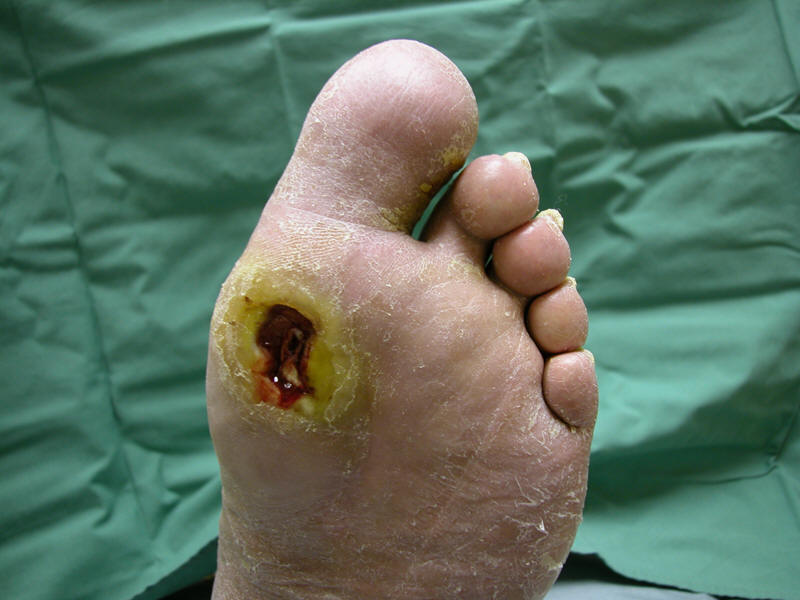
Most diabetic foot infections begin with a break in the protective cutaneous
barrier, typically in the form of a neuropathic ulcer
![]() (I). The lifetime risk of foot
ulceration in persons with diabetes is about 15-25% and about 60% of ulcers are
clinically infected at presentation. A recent prospective study found that
despite patient education and provider follow-up, 9% of diabetic patients
developed a foot infection during a two year observation period; nearly all
of these were precipitated by a foot wound. In developed countries, 85% of lower
extremity amputations in diabetic patients are preceded by a foot ulcer. A
complex, incompletely understood interplay of risk factors may conspire to cause
foot ulceration and foot infection in persons with diabetes (Table 1).
(I). The lifetime risk of foot
ulceration in persons with diabetes is about 15-25% and about 60% of ulcers are
clinically infected at presentation. A recent prospective study found that
despite patient education and provider follow-up, 9% of diabetic patients
developed a foot infection during a two year observation period; nearly all
of these were precipitated by a foot wound. In developed countries, 85% of lower
extremity amputations in diabetic patients are preceded by a foot ulcer. A
complex, incompletely understood interplay of risk factors may conspire to cause
foot ulceration and foot infection in persons with diabetes (Table 1).
Peripheral sensory neuropathy plays the central role in the development of foot ulcers, primarily by causing a loss of protective sensation. Importantly, most patients do not recognize the loss of protective sensation, underscoring the need for preventive efforts including serial surveillance screening and daily foot inspection. Skin ulceration usually results from unperceived repetitive sheer-type trauma due to altered weight bearing (foot deformities and excess plantar pressure), ill fitting shoes, or less commonly, skin-penetrating trauma. Peripheral motor neuropathy can cause abnormal foot biomechanics and lead to distorted foot anatomy. Dry, thickened, and cracked skin related to peripheral autonomic neuropathy increases the risk of skin breaks, offering a portal of entry for bacteria. Most foot ulcers develop on the plantar surface of the foot, especially at the metatarsal heads and to a lesser degree on the toes and calcaneum, the sites of highest pressure with standing and ambulation.
Once a foot ulcer develops, several poorly characterized immunological and
metabolic perturbations may impair healing and allow the infection to progress.
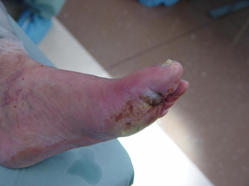
Another risk factor for developing an infection, and for worsening its severity,
is limb ischemia
![]() (II).
Peripheral arterial disease, typically affecting the major arteries between the
knee and ankle, is twice as common in persons with diabetes as in non-diabetics. Diminished tissue perfusion inhibits wound healing and
impairs delivery of antibiotics to infected tissue. Diabetes-related immune
dysfunction, caused by impaired neutrophil and macrophage chemotaxis and
phagocytosis, also predisposes to more frequent, rapidly spreading and severe
infections.
(II).
Peripheral arterial disease, typically affecting the major arteries between the
knee and ankle, is twice as common in persons with diabetes as in non-diabetics. Diminished tissue perfusion inhibits wound healing and
impairs delivery of antibiotics to infected tissue. Diabetes-related immune
dysfunction, caused by impaired neutrophil and macrophage chemotaxis and
phagocytosis, also predisposes to more frequent, rapidly spreading and severe
infections.
|
I. Neuropathic uninfected ulcer |
II. Polymicrobial diabetic foot infection |
|
|
|
Table 1. Risk Factors for Foot Ulceration and Infection
|
Risk factor |
Mechanism leading to ulceration, impaired wound healing or infection |
|
Peripheral sensory neuropathy |
Loss of protective sensation (e.g., repetitive shear-type stress leading to ulceration) |
|
Peripheral motor neuropathy |
Abnormal foot anatomy and biomechanics resulting in excess pressure |
|
Peripheral autonomic neuropathy |
Impaired sweating leading to dry, cracked skin |
|
Arterial insufficiency |
Diminished delivery of nutrient, oxygen, neutrophils, etc. leading to impaired wound healing and clearance of infection |
|
Hyperglycemia |
Immune system (e.g., neutrophil) dysfunction and excess collagen cross-linking |
|
Patient disability or non-adherence |
Reduced vision (unable to inspect feet), prior amputation, lack of regular follow-up with medical care, poor hygiene, inappropriate footwear |
Microbial Etiology
Selecting an appropriate empiric antibiotic regimen requires knowing the usual etiologic organisms (Table 2). Numerous studies have assessed the microbiology of diabetic foot infection, but because of the heterogeneity of the patient populations and culture methods used the reported results vary substantially. Isolation of multiple species of bacteria from a wound specimen is common with many studies reporting a mean of 2-5 isolates per case. Isolation of bacteria from a wound specimen does not define pathogenicity and distinguishing pathogens from colonizers may be difficult. Nonetheless, in almost all cases collecting appropriately obtained specimens for culture is helpful. The results of these cultures may allow tailoring of the antibiotic regimen – either to a narrower spectrum or one targeted to specific bacteria not covered by the initial empiric regimen. Almost all studies, however, show that aerobic gram-positive cocci, particularly Staphylococcus aureus, are responsible for most acute infections, especially in patients who have not recently received antibiotic therapy. Methicillin-resistant Staphylococcus aureus (MRSA), either health-care associated or community-acquired, is an increasingly frequent pathogen in diabetic foot infections. Several studies have reported that a substantial proportion of Staphylococcus aureus are methicillin resistant and that these pathogens may be associated with worse outcomes. Coagulase-negative staphylococci are also frequently isolated, and may be mistakenly dismissed as contaminants. Because persons with diabetes are immunologically compromised, these are often true pathogens. Beta-hemolytic streptococci (usually group B) are also relatively frequent pathogens especially in patients with cellulitis. Enterococci are among the more common isolates in many studies, but their clinical significance is uncertain. Because cephalosporins are active against many common pathogens in diabetic foot infection but not enterococci, treatment with this class predisposes to infection with Enterococcus spp.
Aerobic gram-negative bacilli, usually including Enterobacteriaceae (Escherichia coli, Proteus, Klebsiella, and Enterobacter) are also frequently isolated from diabetic foot infection, especially chronic or previously treated infections. Gram-negative rods are rarely the sole, or even predominant, pathogen. Many broad-spectrum antibiotic agents will cover Enterobacteriaceae, but not Pseudomonas aeroginosa. Pseudomonas deserves specific mention, as it is a relatively frequent isolate and usually requires specifically targeted therapy. This water-borne organism often colonizes, and sometimes infects, wounds that have been soaked or subjected to hydrotherapy. Gram-negative organisms that elaborate extended spectrum beta-lactamases (ESBL) are also a growing problem in diabetic foot infection. As with Pseudomonas, these generally require specifically targeted regimens. Obligately anaerobic organisms may also cause diabetic foot infection. Recognizing these isolates requires obtaining proper (usually tissue) specimens and then quickly and appropriately processing them. The most frequent anaerobic isolates are peptococci and peptostreptococci, and less often Bacteroides species. These may be important pathogens, but almost exclusively occur as part of a polymicrobial infection with an ischemic or necrotic wound. The clinician must also be aware that the causative organism may change over time, especially if the patient’s infection fails to clinically respond to simple regimens.
Table 2. Pathogens Associated with Diabetic Foot Infection Syndromes
|
Diabetic foot infection syndrome |
Pathogen |
|
Cellulitis without ulceration |
Beta-hemolytic streptococci (especially group B) and Staphylococcus aureus |
|
Ulcer or wound, recently developed and no prior antibiotic treatment |
S. aureus and beta-hemolytic streptococci |
|
Ulcer or wound, chronic or recent antibiotic treatment |
Usually polymicrobial – S. aureus and beta-hemolytic streptococci plus Enterobacteriaceae. Enterococci if previous cephalosporin therapy. |
|
Ulcer or wound, prior hydrotherapy or green-blue colored drainage |
Pseudomonas aeroginosa (often in combination with other organisms) |
|
Extensive necrosis or gangrene, ischemic limb, feculent odor (“fetid foot”) |
Polymicrobial – mixed aerobic gram-positive cocci (including enterococci), Enterobacteriaceae, nonfermentative gram-negative rods, and obligate anaerobes |
|
Healthcare-associated |
MRSA; ESBL-producing gram-negative rods |
MRSA = Methicillin-resistant Staphylococcus aureus
ESBL = Extended spectrum beta-lactamase