![]()
Osteomyelitis - Antibiotic Therapy
Principles of Antimicrobial Therapy
As bacteria become more resistant antibiotic therapy can be challenging, therefore an adequate attempt at identifying a microbiologic pathogen is critical. Rarely will rapid initiation of antibiotic therapy change the clinical course, unless the patient is showing significant signs of systemic toxicity. Therefore, appropriate consultation and attempts to obtain bone cultures should be the first treatment. However, there are times when bone cultures will not be possible or cultures will be negative (despite pathologic evidence suggesting osteomyelitis) and therapy will be empiric. Empiric therapy should be guided based on the risk and host factors mentioned in Tables 1 and 3. Previous history of bacteremia (especially with MRSA) should be taken into account due to the potential for bacterial seeding of bone during episodes of bacteremia. When treatment is empirical, clinical response should be evaluated after 2 weeks of treatment to determine if therapy is adequate. Evidence of response would include, but is not limited to, decreased acute phase reactants along with decreased pain, swelling and tenderness. Radiographic changes will be delayed and are not a reliable means of monitoring response to treatment.
Table 1: Microorganisms in Acute Hematogenous Osteomyelitis
Organism Patient Category Staphylococcus aureus
Group B Streptococcus
Escherichia coli
Infants
Staphylococcus aureus
Streptococcus pyogenes
Hemophilus influenzae
Children (up to 4 years of age)
Staphylococcus aureus
21 years or older
Gram negative rods
Elderly
Candida spp.
Patients with intravascular devices or history of candidemia
Staphylococcus aureus Pseudomonas aeruginosa
IV drug abuse
Tuberculosis
HIV, IV drug abuse, alcoholism, homelessness, immunosupression,
history of a positive PPD and former residence in an endemic area
Table 3. Osteomyelitis In A Special Clinical Setting
Risk Factors Organism Site I.V. drug abuse
S. aureus
P. aeruginosa
Serratia
Axial skeleton
Sterno-clavicular joint
Sacro-ileac joint
Hemoglobinopathy
Salmonella
Long bones
Tooth abscess
Anaerobes
Mandible
Diabetic foot abscess
Polymicrobial
Small bones of hands and feet
Human bite
Eikenella Corrodens
Staph Aureus
Hands
Animal Bite
Pasteurella Multocida
Hands, feet, face
Puncture wound of foot
P. aeruginosa
Calcaneus
Median sternotomy
S. aureus
S. epidermidis
Axial skeleton
Meat handlers
Brucella
Flat bones
Fishermen
M. marinum
Small bones of hand
Hemodialysis
S. aureus
S. epidermidis
Axial skeleton
Positive PPD
M. tuberculosis
Thoracic spine
Exposure to TB
Resident in endemic setting
Prosthetic devices
S. aureus
S. epidermidis
Extremities and joints
Antibiotics must be nontoxic, convenient to administer and cost-effective. The selection is based upon in-vitro susceptibilities of the microorganisms causing the infection and ability to penetrate the bone. The therapy is often prolonged requiring outpatient management, administered intravenously for at least the first two weeks following surgery. The regimens for the initial treatment of osteomyelitis are listed in Table 7. Antibiotics should be tailored based on the cultures and the specific needs of each patient. Due pharmacological complexities of the available antibiotics, their adverse effects and possible complications, a dedicated infectious disease specialist often times facilitate the treatment process. Although most of the early reports suggest such an arrangement may improve outcomes, this has yet to be supported by level I evidence-based data.
The duration of antibiotic therapy for osteomyelitis varies as some researchers have recommended as few as two weeks of therapy and others suggested long-term therapy and there is no general consensus. Short-term therapy is indicated in otherwise healthy patients with total debridement and healthy host tissue. The long-term antibiotic therapy is based upon biofilm technology data. These recommendations are typically made for patients requiring reconstruction surgery involving a large volume of graft and of inert materials. These inert materials are the potential site for colonization of bacteria from the blood stream or remnants in the tissue bed until complete revascularization takes place. Selecting the appropriate oral antibiotic and monitoring of antibiotic therapy can minimize bacterial seeding of the reconstruction site. In order to increase the patient compliance antibiotic agents selected must be least toxic, least expensive and require administration once or twice daily. The oral antibiotics with excellent oral bioavailability are listed in Table 8. These antibiotics may be substituted for intravenous agents whenever possible provided that the microorganism is susceptible to these agents.
Because of increased incidences of vancomycin-resistant Enterococcus, especially in intensive care units, and vancomycin-resistant Staphylococcus aureus have been recently reported, vancomycin should be used only if there is high rate of infection caused by methicillin-resistant S. aureus or methicillin-resistant S. epidermidis. A single dose of vancomycin administered before surgery and followed by two or three doses postoperatively should provide adequate peri-operative prophylaxis. Vancomycin should only be used with type 1 hypersensitivity to cephalosporins that includes those with urticaria, laryngeal edema, and bronchospasm with or without cardiovascular shock. Clindamycin is considered a good alternative to cefazolin.
Table 7. Initial Antibiotic Regimens for Patients with Osteomyelitis
Organism
Primary Antibiotic
Alternative Antibiotic
Staphylococcus aureus or coagulase-negative (methicillin-sensitive) staphylococci
Oxacillin or cefazolin
Clindamycin, vancomycin or daptomycin
S. aureus or coagulase-negative (methicillin-resistant) staphylococci vancomycin +/- Rifampin
Daptomycin, Linezolid, trimethoprim-sulfamethoxazole or minocycline plus rifampin
Varied streptococci (groups A and B b-hemolytic organisms or penicillin-sensitive Streptococcus pneumoniae)
IV Penicillin G
Clindamycin, azithromycin, vancomycin or ceftriaxone
Intermediate penicillin-resistant S. pneumoniae
ceftriaxone
Clindamycin, Azithromycin or levofloxacin
Penicillin-resistant S. pneumoniae
vancomycin
levofloxacin
Enterococcus species
Ampicillin or vancomycin
Linezolid, daptomycin
Enteric gram-negative rods
Fluoroquinolone
ceftriaxone
Serratia species
levofloxacin
Ertapenem
Pseudomonas aeruginosa
Cefepime, piperacillin, ciprofloxacin
Imipenem
Anaerobes
Clindamycin
amoxicillin-clavulanate, Ertapenem, moxifloxacin or metronidazole
Mixed aerobic and anaerobic organisms
amoxicillin-clavulanate, 875 mg and 125 mg, respectively, orally BID
Ertapenem, moxifloxacin
Table 8. Selected Oral Antimicrobial Agents with Excellent Oral Bioavailability Commonly Used to Treat Patients with Musculoskeletal Infection
| Antimicrobial Agent |
| Doxycycline Fluoroquinolones Ciprkofloxacin Levofloxacin Moxifloxacin Metronidazole Linezolid Rifampin (must give in conjunction with a second antibiotic) Trimethoprim-sulfamethoxazole |
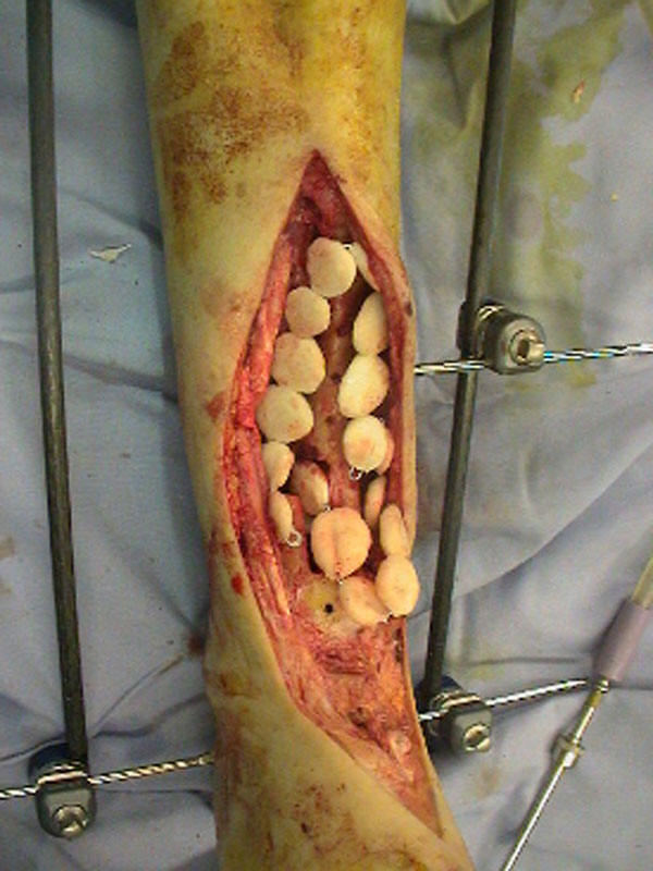
Antibiotic Depot Devices and Techniques
Use of antibiotic depots
![]() allows for high local concentrations of antibiotic with
little systemic absorption Agents have to be heat stable and in powder form. Tobramycin and Vancomycin are the most commonly used antibiotics for depot
delivery. Antibiotic release is bi-phasic, most occurring during the first hours
to days post-implantation and the remaining elution persisting for weeks and
sometimes for years. Some of the other antibiotics that have been tried with
Polymethylmethacralate (PMMA) include clindamycin which elutes well, but is not
available as a pharmaceutical grade powder; Fluoroquinolones in cement powder
has not been reported; Erythromycin is heat stable but demonstrated inadequate
elution from the cement; Tetracycline and colistin fail to elute from the
palacos cement in clinically meaningful quantities. Most of the antibiotic
cement use in the US has been off label use by the surgeon, and despite very
encouraging results from several studies, their approval has been slow. There
are also newer types of material available for local delivery of antibiotics
which are resorbable and do not require removal (Figure 3).
allows for high local concentrations of antibiotic with
little systemic absorption Agents have to be heat stable and in powder form. Tobramycin and Vancomycin are the most commonly used antibiotics for depot
delivery. Antibiotic release is bi-phasic, most occurring during the first hours
to days post-implantation and the remaining elution persisting for weeks and
sometimes for years. Some of the other antibiotics that have been tried with
Polymethylmethacralate (PMMA) include clindamycin which elutes well, but is not
available as a pharmaceutical grade powder; Fluoroquinolones in cement powder
has not been reported; Erythromycin is heat stable but demonstrated inadequate
elution from the cement; Tetracycline and colistin fail to elute from the
palacos cement in clinically meaningful quantities. Most of the antibiotic
cement use in the US has been off label use by the surgeon, and despite very
encouraging results from several studies, their approval has been slow. There
are also newer types of material available for local delivery of antibiotics
which are resorbable and do not require removal (Figure 3).
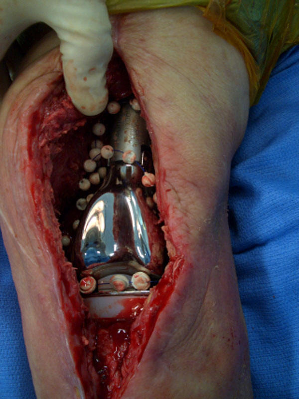
Antibiotic beads inserted into an area of osteomyelitis A prosthetic joint with the presence of antibiotic beads
Chronic Suppressive Therapy
In certain performed (Class C hosts), if the hardware has to be left in place to maintain stability such as an acute infection or if the patient refuses surgical treatment eradication of the infection is unlikely. In such instances, use of chronic suppressive therapy may be in order which typically involves long-term administration of oral antibiotic therapy. Microbiologic diagnosis must be attempted before considering chronic suppressive therapy and the patient must be compliant and be able to tolerate long-term antibiotic therapy. The exact duration of therapy is not clearly defined. However, a six-month course is typically administered to contain and not to eradicate the infection. In patients with fractures suppressive antibiotic therapy is maintained until healing occurs followed by removal of the hardware. In some patients, suppressive antibiotic therapy may be required indefinitely.
PREVENTION
Prevention of osteomyelitis implies decreasing known risk factors in host. For hematogenous osteomyelitis, major risk factors include, indwelling intravascular catheters, distant focus of infection and I.V. drug abuse. For contiguous osteomyelitis, risk factors include trauma, adjacent soft tissue infection, bites and puncture wounds. Patients with diabetes have multiple risk factors inherent to the disease along with vascular insufficiency. Systemic and local co-morbid conditions need to be corrected to avoid osteomyelitis. Smoking cessation and adequate nutrition play an important role in the outcome of osteomyelitis.