
Lymphadenopathy
Authors: Masashi Narita, M.D.
DEFINITION
Lymphadenopathy is an acute or chronic abnormal state of either size or consistency of the lymph nodes; it is a response to infection, inflammation, or malignancy. The body has approximately 600 lymph nodes, but only those in the submandibular, axillary or inguinal regions are normally palpable in healthy individuals. Normal nodes are usually less than 1.0 cm in diameter and tend to decrease or remain stable in size. Lymphadenitis is defined as an acute or chronic inflammation of lymph nodes. Lymphangitis is defined as an inflammation of lymphatic channels, usually in the subcutaneous tissues.
ANATOMY
Lymph nodes are oval bean-shaped structures. Each lymph node is enclosed by a fibrous capsule. Lymph moves into a node via different lymphatic vessels and emerges by one or two efferent vessels. Fibrous septa or trabeculae extend from the covering capsule toward the center of the node. When an infection is present, germinal centers form and the node begins to release lymphocytes. If this barrier of host resistance is overcome, the infectious process may spread to more distant nodes or to the blood stream.
PATHOGENESIS AND PATHOLOGY
The lymph nodes participate in the filtration of microorganisms, the production of antibody, and the processing of lymphocytes within the human body. With the exception of neoplasia, lymph nodes are rarely the site of primary disease. However, they are involved in virtually all infectious processes. When a local infection occurs, the regional lymph nodes react to the invading microorganisms. If this barrier of host resistance is overcome, the infectious process may spread to more distant nodes or to the blood stream. Generalized lymphadenopathy and reticuloendothelial system hyperplasia can occur secondary to widespread sepsis.
Lymph nodes enlarge because of lymphocyte proliferation or macrophage hyperplasia resulting from infiltration of microorganisms or malignant cells, antigenic interaction, or neoplastic proliferation of lymphocytes or phagocytes. Macroscopically, the nodes of acute nonspecific lymphadenitis become swollen, gray-red, and engorged. Histologically, lymphoid follicles are prominent, with large germinal centers containing numerous mitotic figures. When bacteria are the cause of the reaction, the centers of the follicles may undergo necrosis with suppuration. With less severe reactions, there is a neutrophilic infiltrate about the follicles, and numerous neutrophils can be found within the lymphoid sinuses. The cells lining the sinuses become hypertrophied and often undergo hyperplasia.
Acutely inflamed nodes become enlarged because of cellular infiltration and edema. If capsular distension is marked, tenderness may be present. The nodes may become fluctuant if invaded by bacteria. Chronic inflammation can be manifested by enlarged nodes with mononuclear cell infiltration.
Lymphadenopathy can be categorized as (a) acute or chronic, (b) local or generalized, and (c) with or without specific histology. Most patients who present with lymphadenopathies have acute, localized processes, and biopsy of the involved nodes show nonspecific histology, and are classified as regional nonspecific lymphadenitis.
![]()
DIFFERENTIAL DIAGNOSIS
Local Adenopathy
Localized lymphadenopathy can suggest the site of infection, although not necessarily the original source of infection. For example, the sexually transmitted disorders usually affect the genitalia and inguinal lymph nodes, but if the primary lesion (e.g., a syphilitic chancre) is located elsewhere, e.g., the mouth, the adenopathy will be in the cervical area. Similarly, diseases spread by insect vectors will cause adenopathy in the area corresponding to the site of inoculation (Table 1).
Cervical Adenitis
Group A Streptococcus: Cervical adenitis is usually secondary to pharyngitis by respiratory viruses or by group A streptococcus. Group A streptococcal infection (Streptococcus pyogenes) of the pharynx can cause tender anterior cervical adenopathy. The appearance of the pharynx, the presence of exudative material, and the character of the nodes are all nonspecific.
Epstein Barr Virus: Infectious mononucleosis is usually associated with a pharyngitis that may be exudative. Cervical adenitis involving both the anterior and posterior cervical nodes is almost always present. Mononucleosis should be considered as a cause of pharyngitis in the adolescent or young adult. The lymphadenopathy may not be localized to just the cervical region but may be generalized. Splenomegaly, palatal petechiae, supraorbital edema and a generalized maculopapular rash may occur. Hepatomegaly and/or abnormalities of liver function are common in infectious mononucleosis.
Cytomegalovirus: Cytomegalovirus (CMV) infection can mimic infectious mononucleosis, but the Monospot and EBV antibody tests are negative. The pharyngitis and cervical adenopathy in CMV infection is less pronounced than in EBV mononucleosis.
Rarer bacterial etiologies of pharyngitis include Neisseria gonorrhoeae (severe pain but minimal erythema), A. haemolyticum (may be accompanied by a diffuse erythema) and Yersinia species (sometimes suggested by gastrointestinal symptomatology).
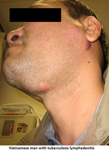
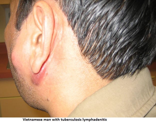
Mycobacterial Species: Scrofula (tuberculous cervical lymphadenitis) presents as painless, enlarged and matted lymphnodes; fluctuation may develop in some nodes. Spontaneous drainage of caseous material onto the skin surface (scrofuloderma) may occur. The node enlargement is usually unilateral and isolated to one group of nodes. The disease is usually located to the lymph nodes only. This syndrome is typically seen in children who are otherwise healthy and have a negative chest x-ray. The responsible pathogens are M. scrofulaceum and M. avium complex (MAC). Mycobacterial cervical adenitis in young adults, aged 20 to 40, is usually caused by classical M. tuberculosis ![]() .
.
Active tuberculosis is usually present elsewhere in the body, although the chest x-ray may show no evidence of tuberculosis in 50 percent of patients. Lymph nodes in mycobacterial infections may be large and matted together and are usually painless. The course of tuberculosis adenitis is generally chronic. Although the caseating granulomas are the classical findings, this is not diagnostic unless the organisms can be identified. Specimens for cultures must be taken at the time of excision or biopsy. A positive tuberculin skin test can suggest the diagnosis. Tuberculosis adenopathy caused by M. tuberculosis usually responds to antituberculosis therapy alone, but excision is generally required to treat disease caused by atypical mycobacteria.
Corynebacterium Diptheriae: Diphtheria is an extremely rare cause of acute cervical lymphadenopathy except in developing countries. History of immunization is crucial. Diphtheria should be suspected in any nonimmunized patient with a pharyngitis accompanied by a gray pseudomembrane that is difficult to remove. Cervical adenopathy and edema of the anterior neck and submandibular areas frequently occur with this form of pharyngitis. Other much more frequent causes of a pharyngeal pseudomembrane are EBV, Streptococcus group A, fusobacterium, Candida albicans, and upper respiratory tract viral pathogens.
Kawasaki’s Disease: The mucocutaneous lymph node syndrome (MLNS) is characterized by protracted fever (longer than five days) associated with cervical adenopathy, conjunctival injection, redness of pharynx and lips, and diffuse scarlatiniform rash including the palms and soles, with subsequent desquamation. The disease may be mistaken for Group A streptococcal scarlet fever, but the diagnostic studies for streptococcal infection are negative and penicillin is ineffective. The fatality rate (about 1%) is related to coronary arteritis.
Kikuchi’s Disease: Kikuchi’s disease (also called Kikuchi-Fujimoto disease or Kikuchi’s histiocytic necrotizing lymphadenitis) occurs predominately in females.Clinical features are characterized by fever, cervical lymphadenopathy, and transient skin rash. Signs and symptoms usually resolve spontaneously within one to four months. The definitive diagnosis of Kikuchi’s disease is made by lymph node biopsy. Histological findings show follicular hyperplasia in early phase, the presence of blast cells in the “proliferative phase”, and necrosis with histiocytes in the “necrotizing phase”. This syndrome is more common in Asia than elsewhere.
Other Nonbacterial Causes: Adenovirus and herpes simplex type 1 can produce severe exudative and ulcerative pharyngitis, as can primary HIV infection. Mycoplasma may cause a mild pharyngitis similar to viral infection. Cervical adenopathy may be mimicked by other inflammatory processes of the neck including parotitis and Ludwig’s angina.
![]()
Occipital Lymphadenopathy
Local infections of the scalp such as pediculosis capitis, impetigo, and ringworm may cause lymphadenopathy in the occipital region. Systemic infections such as syphilis and tuberculosis may enlarge occipital nodes, but usually involve other nodes more dramatically. Rubella virus causes generalized infection that involves occipital nodes; this adenopathy may precede the rubella rash by several days. The nodes, although generalized, usually predominate in occipital, mastoid, and posterior cervical locations. Rubeola also can cause this pattern of adenopathy in addition to rash, fever, and upper respiratory symptoms.
Peripheral Axillary, Epitrochlear, and Inguinal Adenopathy
Enlargement of the axillary and epitrochlear or inguinal nodes occurs most frequently from bacterial infection on the extremities. Repeated trauma to the lower extremities can lead to inguinal lymphadenopathy. Nonspecific inguinal adenopathy is common such that biopsy of the inguinal nodes is usually nondiagnostic. If other nodes are enlarged and accessible, biopsy may be more productive there.
Group A Streptococcus: Group A streptococcal infection of the extremities may produce cellulitis and lymphangitis with regional adenopathy. Fever and superficial red streaks may occur, with painful and tender swollen regional nodes.
Francisella Tularensis: Tularemia of the ulceroglandular type is most frequently associated with the bite of a tick or deerfly, or with contact with the carcass of an infected animal. The site of inoculation is characterized by an ulcerating papule. Regional adenopathy, the location of which depends on the inoculation site, is manifested by hot, large, fluctuant, and tender lymph nodes that may suppurate and drain spontaneously. On the other hand, the node enlargement may at times be much less dramatic and recognized only after very careful physical examination.
Bartonella Species: Cat Scratch Disease may be manifest by localized adenitis in the epitrochlear, axillary, and inguinal areas depending on the site of the scratch or bite. The etiology of cat-scratch disease is Bartonella henselae, a fastidious gram-negative rod. Facial lesions may be associated with cervical or auricular adenopathy. The primary lesion is a tender papule that is frequently capped by a vesicle. Regional nodes then become enlarged and tender, and the overlying skin may be red. If the node becomes fluctuant, it should be aspirated but not incised. Aspiration may reduce local pain with a dramatic lysis of fever.
Yersinia Pestis: The most common form of disease, bubonic plague, is manifested by high fever and systemic toxicity, as well as by an erythematous, edematous matted group of inguinal or axillary nodes that can suppurate and drain spontaneously. The inoculating flea bite is usually not apparent. The enlarged lymph node, the bubo, is extremely tender. The diagnosis can be made by culture of an aspirated lymph node.
Spirillium Minus: Rat-bite fever caused by S. minus may cause rash, lead to the syndrome of a swelling at the site of the bite, regional adenopathy, and fever. The local infected nodes are tender, firm, and freely movable. One to four weeks after the bite, the local lesion flares again as the systemic phase of the illness begins. Fever, generalized rash, and arthritis occur. This infection is more common in Asia and is called Sodoku in Japan. In the other form of rat-bite fever, which is more common in the Western hemisphere is caused by Streptobacillus moniliformis, adenopathy is not prominent.
Sporotrichum Schenkii: Sporotrichosis is often manifest by primary lesion of the upper extremities and the characteristic intermittent hard red lumps along the lymphatics. The nodes then enlarge and the cutaneous nodules may ulcerate. Pain and fever are absent, and the streaking seen in acute bacterial lymphangitis is rare. Sporotrichosis is diagnosed by culture of the nodules, since neither serology nor scrapings or the lesions for staining are helpful. Usually the infection stays localized but requires a long time for resolution. Although rare, primary cutaneous coccidioidomycosis, nocardiosis, and blastomycosis may mimic local sporotrichosis.
Herpes Zoster: Most commonly seen on the trunk, herpes zoster causes a syndrome of clustered vesicles in a unilateral segmental distribution, frequently preceded and accompanied by marked paresthesias, mild fever, and headache. Signs and symptoms usually precede the rash by a few days but at times by as long as one to two weeks. Regional adenitis is a consistent feature of herpes zoster. The nodes are nontender and freely movable.
Herpes Simplex (Herpetic whitlow): Epitrochlear and axillary adenopathy may be a complication of herpetic whitlow, an infection of the pulp of a finger caused by Herpes simplex, usually type 1. This primary lesion is usually very painful.
Rickettsioses: Rickettsial infection present with fever, headache, myalgic and regional lymphadenopathy. The etiologies include Oritentia tsutsugamushi (Scrub Typhus), Rickettsia akari (Rickettsialpox), and other spotted fever group Rickettsial infections including R. australis (Queensland tick typhus), R. honei (Flinders Island spotted fever), R. conorii (African tick bite fever), R. sibirica (Siberian tick typhus), R. sibirica mongolotimonae (Lymphangitis-associated rickettsiosis), R. slovaca (Tick-borne lymphadenopathy).
![]()
Inguinal Adenopathy
Many sexually transmitted infections are responsible for inguinal lymph node enlargement. Frequently, a genital ulcer accompanies the adenopathy, producing the “ulcer-adenopathy” syndrome.
Treponema Pallidum: Syphilis often presents as a painless skin lesion, the chancre, usually on the external genitalia, with enlarged freely movable, firm, nonsuppurating, and painless inguinal nodes. The chancre heals in two to six weeks, but the lymphadenopathy may persist for months. Diagnosis of primary syphilis is made by a characteristic lesion and serology.
Haemophilus Ducreyi: Unlike syphilis, the genital ulcers and inguinal nodes of chancroid are tender and painful. If untreated, the nodes may suppurate and rupture to form a large single ulcer. This last complication may be prevented by aspiration when the node is tense.
Lymphogranuloma Venereum (LGV): Chlamydia trachomatis is the cause of Lymphogranuloma vereum, a genital ulcer disease. Inguinal adenopathy is prominent in LGV and may progress to multiocular suppuration with multiple fistulae. At times the inguinal ligament forms a line of cleavage through the matted nodes, which are enlarged both above and below the ligament (the “Groove” sign). The primary genital lesion is a single small ulceration that generally goes unnoticed. It has usually disappeared by the time the lymph node enlargement occurs. LGV may be accompanied by generalized constitutional symptoms and is occasionally associated with severe arthralgias, diarrhea, and hyperglobulinemia. It is rare in developing countries but can be endemic in tropical countries. PCR of specimens taken from the ulcer is now the most sensitive diagnostic method.
Klebsiella Granulomatosis: Klebsiella granulomatous is the cause of granuloma inguinale (Donovanosis). The disease is manifested by a painless genital papule anad nodule that erodes to leave a beefy red granular base. The infection spreads to the inguinal area and produces a marked subcutaneous swelling. The lesion is called a “pseudobubo” since it is not really due to lymph node involvement, but rather to soft tissue granulation and frequently ulceration. This infection is rare in developing countries.
Herpes Viruses: Genital herpetic lesions are characteristically multiple, small erosions on an erythematous base, typically arranged in a cluster. They are extremely painful and are often accompanied by bilateral or unilateral inguinal adenopathy.
Generalized Adenopathy
Fever and generalized adenopathy are usually due to viruses, especially EBV and rubella, and HIV. EBV mononucleosis may also be mimicked by infection with CMV, Toxoplasmosis gondii and HIV. Acquired or reactivated CMV infection is most frequent in normal young people, in patients who have had surgery with bypass (postperfusion syndrome), recipients of blood transfusions, leukocyte infusions and transplants, and in immunocompromised individuals. Patients with this CMV syndrome may present with protracted fever and with moderate hepatic inflammation along with lymphocytosis and atypical lymphocytes, which occur late in the course of the disease. Splenomegaly and adenopathy are not always present. The hepatitis viruses and HHV-6, the cause of roseola infantium, can also produce lymphadenopathy and a mononucleosis-like illness. Other viruses, particularly rubella, adenovirus, and dengue can also cause significant generalized lymph node enlargement (Table 1).
Primary HIV infection causes a mononucleosis-like illness often accompanied by a diffuse salmon-colored rash that includes the palms and soles. Lymphadenopathy occurs in 50 to 75 % of patients who develop an acute illness approximately 3 to 6 weeks after initial exposure to HIV. In the latency period, persistent generalized lymphadenopathy (PGL) affects between 50 to 70 percent of HIV patients. The lymph nodes are non tender and affect the nodes in the cervical, mandible, inquinal and axillary areas.
Lymphadenopathy is found in 5% of acute viral hepatitis. HBV infection in childhood can be associated with papular dermatitis with enlarged lymph nodes in the axillary and inguinal regions-the Gianotti-Crosti syndrome. This syndrome can also occur after infection by EBV, HAV, HCV, CMV, enterovirus, parvovirus,parainfluenzavirus, rotavirus, respiratory syncytial virus, HIV, HHV-6, Mycoplasma pneumoniae, beta-hemolytic streptococci, Bartonella henselae, Borrelia burgdorferi, and following vaccination with influenza virus vaccine, measles-mumps-rubella vaccine, hepatitis B vaccine, oral polio vaccine or Japanese encephalitis vaccine (1).
![]()
Toxoplasma gondii
The most common syndrome seen in acquired symptomatic toxoplasmosis resembles EBV mononucleosis, with enlarged nonsuppurative lymph nodes, fever, and maculopapular rash. The nodes in toxoplasmosis are firm, discreet, freely movable, and at times, painful and tender. The disease has occasionally been misdiagnosed as Hodgkin’s disease. Frequently the cervical nodes are the largest, increasing the clinical similarity of this entity to mononucleosis. Toxoplasmosis can involve the CNS, liver, heart, lungs, or eyes as target organs. This infection must be considered in patients with an underlying immunosuppressive disease.
Brucella Species
Brucellosis is characterized by fever, chills, hepatosplenomegaly, arthralgia, and leukopenia with relative lymphocytosis. Sarcoiliitis and spondylitis are common. Small, nontender, cervical and axillary node enlargements are common. These nodes may show granulomas or a striking reticuloendothelial hyperplasia with some multinucleate giant cells. Testicular swelling and/or pain may occur.
Histoplasmosis and fungi
Disseminated histoplasmosis and other deep fungal infections are capable of producing widespread lymphadenopathy as part of the generalized reticuloendothelial reaction to these agents.
Treponema pallidum
Secondary syphilis is recognized by nontender, generalized lymphadenopathy with fever and a diffuse nonpruritic maculopapular rash, which often includes the palms and soles. The nodes may remain palpable even months after the disappearance of other stigmata of secondary syphilis, i.e., fever, rash, condylomata, and mucous patches. Pre and postauricular, occipital and epitrochlear nodes are all frequently enlarged in secondary syphilis.
Mycobacterium tuberculosis
Miliary tuberculosis may cause generalized adenopathy. The typical military pattern on chest x-ray may often be delayed.
![]()
Non-Infectious Causes
Many noninfectious disorders include adenopathy among their manifestations, e.g., lymphoreticular malignancies, collagen vascular diseases, and sarcoidosis (Table 2). Peripheral non-tender, lymphadenopathy is common in sarcoidosis; the cervical, axillary, epitrochlear, and inguinal nodes are the most commonly involved regions. The nodes are non-ulcerative unlike infectious causes.
HISTORY
The history in a patient with lymphadenopathy should focus upon demographic information (age, sex, race, ethnicity, occupation, location of residence), the clinical course (acute, subacute or chronic), constitutional symptoms (fever, night sweats, fatigue, weight loss, sore throat, cough), exposure history (pets, medications, infectious diseases), high-risk behavior (sexual behavior and drug abuse), family history (malignancy, tuberculosis and others).
PHYSICAL EXAMINATION
If lymph nodes are palpated, the following five characteristics should be noted and documented in the history: size, tenderness, consistency, location, and fixation. Abnormal nodes are generally greater than 1cm in diameter. In general, normal lymph nodes in the axillary and cervical regions up to 1 cm. in size, in the inguinal regions up to 1.5 cm in size, and the epitrochlear location up to 0.5 cm in size.
A lymph node size of 2.25 cm2 (1.5 x 1.5cm) was the best discriminating limit for distinguishing malignant or granulomatous lymphadenopathy from other causes of lymphadenopathy (11). No patient with a lymph node smaller than 1 cm, 2 had cancer, compared with 8% and 38% of those with nodes 1 to 2.25 cm2 and greater than 2.25 cm2, respectively (11).
Hard nodes with fibrosis are usually associated with malignancy. Firm, rubbery nodes are found in lymphomas and chronic leukemia; nodes in acute leukemia tend to be softer (7). Tenderness occurs when the capsule is stretched during rapid enlargement, usually secondary to an on inflammatory process. Normal lymph nodes are freely movable in the subcutaneous space. Abnormal nodes can become fixed to adjacent tissues by invading cancers or inflammation in tissue surrounding the nodes.
Localized lymphadenopathy should prompt a search for pathology in the area of node drainage, although some systemic diseases can present with local adenopathy (Table 3). Generalized adenopathy is usually a manifestation of systemic disease.
![]()
LABORATORY TESTING
Laboratory testing can be used to confirm a diagnosis that is suspected on the basis of the history and physical examination.
General
Patients with generalized lymphadenopathy should have a CBC and chest radiograph. If these are normal, other test considerations include HIV antibody determination, RPR, ANA, and Monospot test, although these are often of low yield in the absence of a more specific indication (7).
Serological Tests
Serological testing is useful for detection of antibodies for viral illness including EBV, CMV, measles, rubella, dengue and bacterial infections including Yersinosis,Cat Scratch Disease, tularemia, toxoplasmosis, and syphilis. For the diagnosis of acute infectious mononucleosis, the sensitivity of the heterophile test (Monospot) is 90% (5). Antibody to viral capsid antigen (VCA) and early antigen in diffuse pattern (EA-D) should be obtained for suspected cases of acute infectious mononucleosis. The VCA-EBV, IgM is the most sensitive test for acute EBV infection. Titers of IgM and IgG antibodies to VCA are elevated in more than 90% of patients at the onset of disease; the IgM antibodies are absent by 4-8 weeks following acute infection.
In the differential diagnosis of infectious mononucleosis, serology for hepatitis viruses (HBsAg, IgM anti HAV, IgM anti HBc, and anti-HCV), HIV, CMV, and toxoplasma should be ordered. For the diagnosis of primary syphilis, non-treponemal and treponemal tests are supportive (sensitivity 78% and 84%, respectively). The standard screening test for HIV infection is the ELISA with a sensitivity of > 99.5%. The most commonly used confirmatory test is the western blot. Serological tests are not reliable for rat-bite fever, sporotrichosis, lymphgranulmoa venereum (Chlamydia trachomatis).
Microbiological
Fluctuant lymph nodes should be aspirated, while suppurative, ulcerative nodes should be biopsied for culture and microscopic evaluation. Characteristic findings of direct microscopic examination for lymphadenopathy includes intracellular gram-negative diplococci for Neisseria gonorrhoeae, “Chinese characters” on gram stain forCorynebacterium diphtheriae, bacilli in chains or clumps on Warthin-Starry Silver stains for Bartonella henselae, “safety-pin” shaped organisms on Wayson’s stain forYersinia pestis, Giemsa, Wayson and Gram stains for Streptobacillus moniliformis (rat-bite fever), “cigar-shaped organisms” on silver or PAS stain of histopathology forSporotrichosis schenckii, multinucleated giant cell on Tzanck smear for HSV and VZV, Treponema pallidum by darkfield microscopy for primary syphilis, dark staining “Donovan bodies” on tissue crush preparation or biopsy for Klebsiella granulomatis (granuloma inguinale; donovanosis) (Table 4).
Special culture media for fastidious organisms includes Loffler’s or Tindale’s media for Corynebacterium diphtheriae, Cefsulodin-Irgasan-Novobiocin (CIN) agar for Yersinia spp., Thayer-Martin media for Neisseria gonorrhoeae, Cysteine-enriched media for Francisella tularensis, Sabouraud’s agar for fungi (Sporotrichosis,Histoplasmosis), and cell culture or mouse inoculation for Toxoplasma gondii. Primary cutaneous coccidioidomycosis, nocardiosis, and blastomycosis may be diagnosed by biopsy with appropriate stains and cultures. Sporotrichosis is definitively diagnosed by culture of the nodules, since neither serology nor scrapings of the lesions of staining are especially helpful.
Tests for M. tuberculosis
A positive tuberculin skin test, although suggestive, is not diagnostic of tuberculosis because a positive test can occur in individuals without active disease. Moreover, a negative tuberculin skin test may be observed in active tuberculosis (15-25% in tuberculosis lymphadenitis). A PPD may assist in differentiating tuberculosis from sarcoidosis when the lymph node histology is nonspecific. Tuberculin skin is the first step in the diagnosis of cervicofacial lymphadenitis in children without a history of tuberculosis exposure or BCG vaccination. Among total 112 nontuberculous mycobacterial infections (83 of Mycobacterium avium, 21 of Mycobacterium haemophilum, and 8 of other nontuberculous mycobacterial species); tuberculin skin testing had a sensitivity and specificity of 70% and 98%, respectively, and a positive predictive value and a negative predictive value of 98% and 64% at the optimal cutoff for a positive test (5mm) (9).
The QuantiFERON®-TB Gold test (QFT-G) is a whole-blood test for use in diagnosing Mycobacterium tuberculosis infection, including latent tuberculosis infection (LTBI) and tuberculosis (TB) disease. Results are not affected by exposure to BCG or other mycobacterial species. If the patient is infected with M. tuberculosis, their white blood cells will release IFN-gamma in response to contact with the TB antigens. The QFT-G results are based on the amount of IFN-gamma that is released in response to the antigens. Results are reported as positive, negative or indeterminate and do not require the subjective evaluation of reading a PPD. Positive results do not vary based on patient risk factors as do the PPD. Caution must be employed when interpreting results of QFT-G in immunocompromised patients since data is lacking.
![]()
Imaging Studies
Chest radiograph, although seldom positive, should be performed in every patient because it may show the presence of mediastinal lymph node enlargement observed in malignant lymphomas, sarcoidosis, tuberculosis, histoplasmosis, and metastatic cancer. Chest radiograph may also disclose parenchymal lesions compatible with granulomatous fungal diseases, tuberculosis sarcoidosis, metastatic tumors, or lymphomas (11). The computed tomography (CT) and ultrasonography of the involved area may be useful to differentiate lymphadenopathy from other nonlymphatic enlargements, to show lymph node enlargement of the mediastinum or abdomen (11), and to determine the exact anatomic location of the enlarged node before its surgical excision (particularly in the cervical area).
Invasive Diagnostic Methods
Lymph node may be biopsied by excisional methods or needle aspiration. Although fine-needle aspiration is simple, safe, and inexpensive, lymph node disorders such as reactive hyperplasia, Hodgkin disease, and non-Hodgkin lymphoma may be extremely difficult to distinguish from one another with fine-needle aspiration alone. In patients without an established diagnosis and with accessible peripheral adenopathy, excisional biopsy is preferred over fine-needle aspiration to ensure adequate sampling (8).
The changes of chronic inflammation seen on lymph node biopsy may also be nonspecific. Even the presence of epitheloid granulomatous with caseous necrosis and giant-cell formation is not in itself diagnostic of tuberculosis, since the same findings may be seen in tularemia, LGV, cat scratch disease, brucellosis, and even sarcoidosis. With appropriate cultures and stains such as Ziehl-Neelsen, PAS, and the Gomori silver stain, a specific microorganism may occasionally be identified (12).
Increased age (>40 years) suggests malignancy. At younger ages, individuals are more likely to have nonspecific lymphadenitis, infectious mononucleosis, rubella, and toxoplasmosis. Lymph node size is generally determined by palpation and external measurement. It should be conceded that this parameter is not easily measured or reproducible by physical examination. Location is pertinent. Palpable lymph nodes in the cervical axillary and inguinal areas are often observed in normal individuals. In one study, 56% of patients examined for other causes had palpable cervical nodes (10,14). In another study of individuals older than 13 years, palpable nodes were found in the cervical (80%), axillary (70%), and inguinal (90%) areas (15). On the other hand, supraclavicular lymphadenopathy is considered suggestive of serious disease (14). Tenderness or painful lymph nodes are usually benign, while painless hard nodes can be malignant (14). Duration of lymphadenopathy is also useful. A duration of several days is usually infection. Interestingly, a long history (>1 year) is also suggestive of a benign condition (although tuberculosis and lymphoma can have a prolonged course). Duration proved not to be a useful parameter in assessing the lymph node biopsy (14).
In summary, we recommend that the following studies be performed for most patients in whom lymph node biopsy is considered: 1) Monospot test and VCA (viral capsid antigen)-EBV, IgM. 2) Serology for toxoplasmosis. 3) Quantiferon-TB Gold test or PPD skin test for tuberculosis, and 4) chest radiograph to search for pulmonary infiltrates and mediastinal lymphadenopathy.
Clinical parameters favoring malignancy and are increased age (>40 years), large size of the lymph node (>4.0 cm2), supraclavicular location, and hardness of the node. The presence of constitutional symptoms (fever, weight loss, night sweats) is suggestive of a lymphoma or tuberculosis. Excisional biopsy may be warranted.
In general, lymph nodes that have been present outside the inguinal region for longer than 1 month and measure 1×1 cm or larger without an obvious diagnosis should be considered for biopsy (8). The purpose of the biopsy is to rule out malignancy or treatable infectious diseases, especially tuberculosis and disseminated fungal disease. Most patients who present with lymphadenopathies have acute, localized process. Invasive evaluation of these nodes is not generally warranted. The biopsy of these involved nodes shows nonspecific histology, i.e., regional nonspecific lymphadenitis (12). If the underlying cause of lymphadenopathy is suspected to be a viral infection, lymph node biopsy can be deferred.
Prediction Models
Clinical prediction rules have been developed that use key elements of history and physical examination to predict the utility of lymph node biopsy. Centor’s criteria is a simple 4-item clinical prediction rule used to predict the probability of Group A streptococcus infection. Tonsillar exudate, swollen tender anterior cervical nodes, absence of cough, and a history of fever are the key parameters. The rule is accurate, with an area under the receiver operating characteristic (ROC) curve of 0.79. The presence of 3 or 4 findings increases the probability of streptococcal pharyngitis (4).
A mathematical model has been formulated for the prediction of the need of lymph node biopsy develop based on the medical history (age) and the physical findings. The lymph node parameters were tenderness and size of the affected lymph node, supraclavicular location and texture of the lymph nodes (soft, semi-hard, hard). Surprisingly, generalized pruritus was one of the parameters; presence of pruritis correlated with presence of lymphoma, especially Hodgkins disease (14). This model was developed in a university hospital in Greece. A study in a Japanese hospital showed that the results of a model developed in Greece could not readily be extrapolated elsewhere. In the Japanese study (13), tuberculosis and toxoplasmosis were extremely rare as compared to the Greece study. In contrast, Kikuchi’s lymphadenopathy was common in Japan, but rare in Greece. This disparity illustrates how the differences in epidemiology and patient population will affect the incidence of the diverse causes of lymphadenopathy.
![]()
EMPIRIC ANTIBIOTICS
Antibiotics are not indicated for lymphadenopathy unless there is unusually strong evidence of a bacterial infection and the patient is toxic. Glucocorticosteroids should not be used to treat lymphadenopathy because their lympholytic effect was obscure some diagnoses (lymphoma, leukemia, Castleman's disease). Moreover, corticosteroids can contribute to delayed healing or activation of underlying infections. An exception is the life-threatening pharyngeal obstruction by enlarged lymphoid tissue in Waldeyer’s ring that is occasionally seen in infectious mononucleosis (2).
ENDPOINTS FOR MONITORING
Most patients with lymphadenopathy do not require a biopsy. If the patient's history and physical findings point to a benign cause for lymphadenopathy, then careful follow-up at a 2- to 4-week interval can be employed. The watch-and-wait policy is generally applied when lymph node enlargement may be caused by a viral infection for which no specific therapy is available. Close follow-up of the patient at monthly intervals, or earlier if the size of the enlargement is increasing, is necessary.
![]()
REFERENCES
1. Chuh ATC V. Gianotti-Crosti syndrome (papular acrodermatitis): UpToDate, 2007.
2. Braunwald EK, Dennis; Fauci, Anthony; Hauser, Stephen; Longo, Dan; Jameson, J. Harrison's Principles of Internal Medicine: McGraw-Hill, 2004.
3. Brown JR, Skarin AT. Clinical mimics of lymphoma. Oncologist 2004; 9:406-16.[PubMed]
4. Ebell MH, Smith MA, Barry HC, Ives K, Carey M. The rational clinical examination. Does this patient have strep throat? JAMA 2000; 284:2912-8.[PubMed]
5. Farhat SE, Finn S, Chua R, et al. Rapid detection of infectious mononucleosis-associated heterophile antibodies by a novel immunochromatographic assay and a latex agglutination test. J Clin Microbiol 1993; 31:1597-600.[PubMed]
6. Ferrer R. Lymphadenopathy: differential diagnosis and evaluation. Am Fam Physician 1998; 58:1313-20. [PubMed]
7. Fletcher R. Evaluation of peripheral lymphadenopathy in adults: UpToDate Ver 15.1, 2007.
8. Habermann TM, Steensma DP. Lymphadenopathy. Mayo Clin Proc 2000; 75:723-32. [PubMed]
9. Lindeboom JA, Kuijper EJ, Prins JM, Bruijnesteijn van Coppenraet ES, Lindeboom R. Tuberculin skin testing is useful in the screening for nontuberculous mycobacterial cervicofacial lymphadenitis in children. Clin Infect Dis 2006; 43:1547-51. [PubMed]
10. Linet OI, Metzler C. Practical ENT. Incidence of palpable cervical nodes in adults. Postgrad Med 1977; 62:210-3.[PubMed]
11. Pangalis GA, Vassilakopoulos TP, Boussiotis VA, Fessas P. Clinical approach to lymphadenopathy. Semin Oncol 1993; 20:570-82. [PubMed]
12. Schlossberg D. Differential Diagnosis of Infectious Diseases: Williams & Wilkins, 1996.
13. Tokuda Y, Kishaba Y, Kato J, Nakazato N. Assessing the validity of a model to identify patients for lymph node biopsy. Medicine (Baltimore) 2003; 82:414-8. [PubMed]
14. Vassilakopoulos TP, Pangalis GA. Application of a prediction rule to select which patients presenting with lymphadenopathy should undergo a lymph node biopsy. Medicine (Baltimore) 2000; 79:338-47.[PubMed]
15. Zuelzer WW, Kaplan J. The child with lymphadenopathy. Semin Hematol 1975; 12:323-34.[PubMed]
Table 1: Localized and Generalized Lymphadenopathy Disease
| Localized |
|---|
| Cervical adenopathy : |
|
| Occipital adenopathy : |
Nonspecific scalp infection |
| Peripheral adenopathy : |
Location varies with inoculation site: Usually axillary or epitrochlear: |
| Inguinal : |
Primary syphilis |
| General adenopathy |
Infectious mononucleosis (EBV), Rubella, CMV, HIV, Hepatitis virus, Measles, Dengue, Toxoplasmosis, Tularemia (Franciscella tularensis), Brucella, Histoplasmosis, Secondary syphilis, Miliary tuberculosis |
Table 2: Infections Causing Lymphadenopathy Infections
| Infections |
|---|
| Viral : |
| Infectious mononucleosis (Epstein-Barr virus) Cytomegalovirus Infectious hepatitis Adenovirus Herpes simplex, Herpes zoster HHV-6 (Human herpesvirus-6) HIV Human T-lymphotropic virus Rubella Rubeola |
| Bacterial : |
| Cutaneous infections (Staphylococcus, Streptococcus) Cat-scratch fever Chancroid Melioidosis Tuberculosis Atypical Mycobacteria Primary and secondary syphilis |
| Chlamydial : |
| Lymphogranuloma venereum |
| Protozoan : |
| Toxoplasmosis |
| Mycotic |
| Histoplasmosis Coccidiodomycosis |
| Rickettial : |
| Scrub typhus |
| Helminthic : |
| Filariasis |
| Noninfectious Causes |
| Autoimmune : |
| Rheumatoid arthritis Juvenile rheumatoid arthritis Systemic lupus erythematosus Dermatomyositis Mixed connective tissue disease Sjogren’s syndrome |
| Iatrogenic hypersensitivity : |
| Serum sickness Drug hypersensitivity Anticonvulsants: Phenytoin, carbamazepine, primidone Antibiotics: Sulfas, penicillins, cephalosporines, erythromycin Anti-inflammatory : Gold, sulindac, sulfasalazine Others : Aspirin, captopril atenolol quinidine, allopurinol pyrimethamine |
| Neoplasm : |
| Lymphoreticular malignancies Paraproteinemias (monoclonal gammopathy) Metastatic diseases |
| Miscellaneous : |
| Sarcoidosis Amyloidosis Vaccine-induced Kikuchi's disease Hyperthyroidism Storage diseases (Gaucher, Niemann-Pick, Fabry, and Tangier) Severe hypertriglyceridemia Kimura’s disease Kawasaki disease Silicone injection Graft vs host disease Autoimmune hemolytic anemia Gianotti-Crosti syndrome |
Table 3: Lymph Node: Location, Lymphatic Drainage and Selected Differential Diagnosis
| Location | Lymphatic drainage | Causes |
|---|---|---|
Submandibular |
tongue, submaxillary gland, lips, and mouth, conjunctivae |
Infections of head, neck, sinuses, ears, eyes, scalp, pharynx |
Submental |
Lower lip, floor of mouth, tip of tongue, skin of cheek |
Mononucleosis syndromes, Epstein- Barr virus, |
Jugular |
Tongue, tonsil, pinna, parotid |
Pharyngitis organisms, rubella |
Posterior cervical |
Scalp and neck, skin of arms and pectorals, thorax, cervical and axillary nodes |
Tuberculosis, lymphoma, head and neck malignancy |
Suboccipital |
Scalp and head |
Local infection |
Postauricular |
External auditory meatus, pinna, scalp |
Local infection |
Preauricular |
Eyelids and conjunctivae, temporal region, pinna |
Local infection |
Right supraclavicular node |
Mediastinum, lungs, esophagus |
Lung, retroperitoneal or gastrointestinal cancer |
Left supraclavicular |
Thorax, abdomen via thoracic duct |
Lymphoma, thoracic or retroperitoneal cancer, node bacterial or fungal infection |
Axillary |
Arm, thoracic wall, breast |
Infections, cat-scratch disease, lymphoma, breast cancer, bacterial or fungal infection |
Epitrochlear |
Ulnar aspect of forearm and hand |
Infections, lymphoma, sarcoidosis, tularemia, |
Inguinal |
Penis, scrotum, vulva, vagina, perinerum, gluteal Region, lower abdominal wall, lower anal canal |
Infections of the leg or foot, STDs, pelvic malignancy, bubonic plague |
Table 4. Microbiological Findings of Lymphadenopathy
| Organisms and disease | Microscopic | Characteristic findings | Culture media |
|---|---|---|---|
|
(Gonorrhoea) |
Gram stain |
Gram-negative diplococcal "kidney beans" shape |
Thayer-Martin media |
|
(Diphtheria) |
Gram stain |
Gram-positive rods" Chinese character" |
Loffler’s or Tindale’s media |
|
(Cat-scratch disease) |
Warthin-Starry Silver stain |
Bacilli in chains or clumps |
Cultures are not recommended. Low sensitivity: 20% compared with PCR |
|
(Bubonic plague) |
Wayson's stain |
Light blue bacilli with dark blue polar bodies "Safety-pin" shaped organism gram-negative coccobacilli |
Brain-heart infusion, sheep blood agar, chocolate agar, or MacConkey agar
|
|
|
Cefsulodin-Irgasan-Novobiocin (CIN) agar |
|
Fransicella tularensis (Tularemia) |
Gram stain |
Faintly staining, tiny, intracellular and extracellular gram-negative bacilli. |
Cysteine-enriched media |
Streptobacillus moniliformis (Rat-bite fever) |
Gimsa, Wayson's and gram stain |
Pleomorphic bacillary, gram-negative bacilli |
Enriched media (micro-aerophilic medium with increased CO2 ). |
Sporotrichosis schenckii(Sporotrichosis) |
Silver or PAS stain |
"Cigar" shaped organism |
Sabouraud’s agar |
|
|
Sabouraud’s agar |
|
Tzanck smear |
Multinucleated giant cell |
|
|
|
(Primary syphilis) |
Darkfield examination |
A corkscrew appearance of spirochetes and moves in a spiraling motion with a characteristic 90 degree undulation about its midpoint
|
|
Tissue cruch preparation biopsy Wright's or Giemsa's stain |
Dark-staining "Donovan bodies": clusters of blue rods, with prominent polar granules and surrounded by pink capsules, are seen within infected epithelial cells |
Difficult
|
|
|
|
Cell culture or mouse inoculation |
What's New?
Choi P, et al. Polymerase chain reaction for pathogen identification in persistent pediatric cervical lymphadenitis. . Arch Otolaryngol Head Neck Surg. 2009 Mar;135:243-8.
Angelaskis E, et al. Real-time PCR Strategy and Detection of Bacterial Agents of Lymphadenitis. Eur J Clin Microbiol Infect Dis 2009;28:1363-1368.
GUIDED MEDLINE SEARCH FOR
Review Articles
Manuel O, Humar A. EBV-Associated Post-Transplant Lymphoproliferative Diseases in Transplant Recipients - Infectious Disease and Antimicrobial Agents
Aberg JA, et al. Primary Care Guidelines for the Management of Persons Infected with Human Immunodeficiency Virus: 2009 Update by the HIV Medicine Association of the Infectious Diseases Society of America. Clin Infect Dis 2009;49:651-681.