
Why Should I Subscribe?
Antimicrobe is the most comprehensive authoritative database for infectious diseases and antimicrobial agents today. The website is updated daily with new articles on diagnosis and therapy . Alternative drugs, when the drug of choice cannot be used, is a major feature. Reviews, Guidelines, History of Medicine stories and focused Medline searches within the website characterize this innovative and sophisticated approach to medical information.
![]()
![]()
Registered Users Start Here
Take a Look Inside!
View the Table of Contents from Our References.
Click an image below to browse the Table of Contents for the selected book.
-
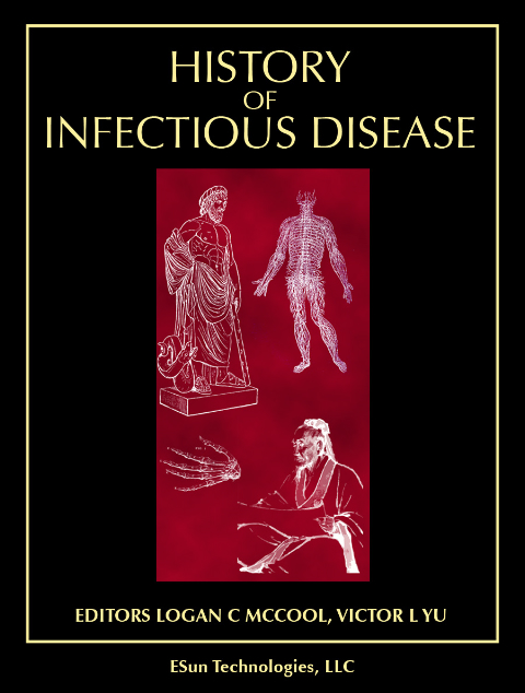
The history of infectious diseases is spread around each monograph under "History" section"
Over 500 international authorities in infectious diseases, epidemiology and pharmacology have contributed to our extensive online database.
![]()
![]()