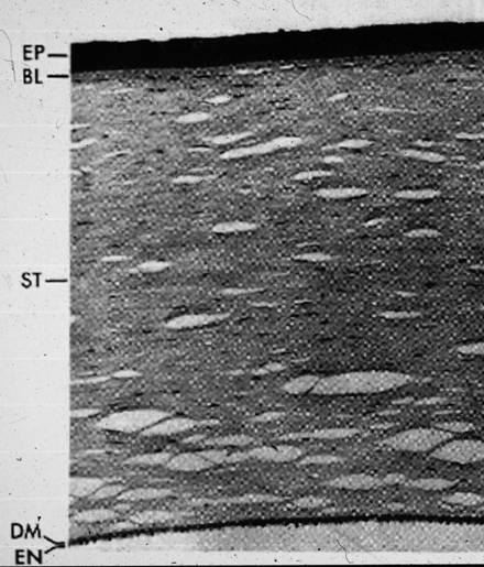
Figure 1. Normal cornea consists of 5 distinct layers. Epithelium (EP), Bowman’s layer (BL), Stroma (ST),
Descemet’s membrane (DM), and Endothelial (EN).
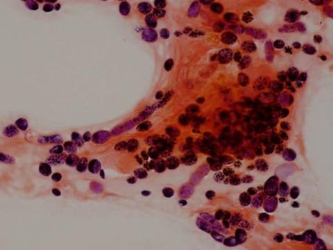
Figure 2. Corneal button of a patient with fungal keratitis.
Note the fungal elements and inflammatory processes in the cornea.
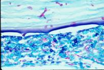
Figure 3. Fungal corneal ulcer.
Stromal infiltration with feathery borders.
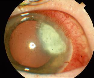
Figure 4. 55 year old horsebreeder and contact lens wearer presented with fungal corneal ulcer.
Note endothelial plaque on slit beam image.
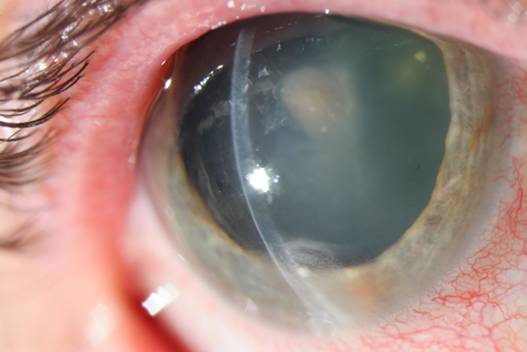
Figure 5: Fungal gram stain of the same patient as figure 4 with Phaeoannellonyces
werneckii keratitis; infection resolved with topical Amphotericin.