Listeria monocytogenes

Major pathogen in CSF and Blood only

@ Ellen Jo Baron 2007

  Motility:

  Tumbling motility at 26°C

  Non-motile at 35° C

Beta hemolysis (β-hemolysis): Beta hemolysis is the complete lysis of the red blood cells around and under the colonies on a blood agar plate. This area appears transparent. Streptococcus pyogenes displays beta hemolysis and is often called Group A beta-hemolytic strep (GABHS).

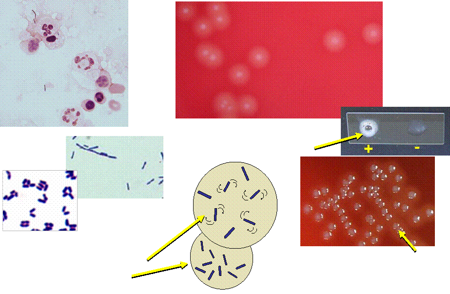
  Beta hemolysis does NOT extend

  beyond edge of colony

Beta hemolysis (β-hemolysis): Beta hemolysis is the complete lysis of the red blood cells around and under the colonies on a blood agar plate. This area appears transparent. Streptococcus pyogenes displays beta hemolysis and is often called Group A beta-hemolytic strep (GABHS).

  This organism is NOT Listeria

  because beta hemolysis extends

  beyond edge of colony

  Regular Gram + rods

  May be short or longer

Gram stain: The Gram stain, is a laboratory staining technique that distinguishes between two groups of bacteria that have differences in the structure of their cell walls. Standard bacterial taxonomy makes a distinction between Gram-negative bacteria, which stain red/pink and the Gram-positive bacteria, which stain blue/purple. Different antimicrobial agents are directed specifically at gram-positive bacteria and gram-negative bacteria.
Catalase test: The catalase test is used to differentiate some bacterial species. The test is done by placing a drop of hydrogen peroxide on a microscope slide. Using an applicator stick, a small portion of a colony is then added to a drop of hydrogen peroxide drop.

  Colony looks like Group B streptococci.

  Differentiate from Group B strep by Gram stain

  and positive catalase reaction

CSF showing PMNs,  Monocytes, &

 Gram positive rods (may be intracellular)