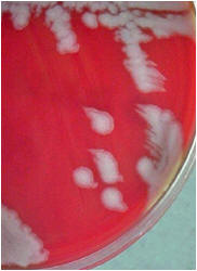
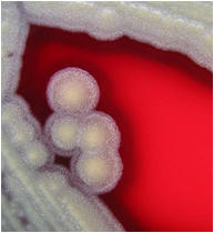
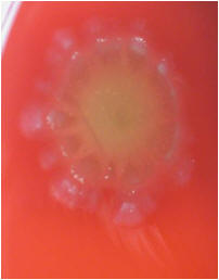
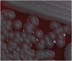
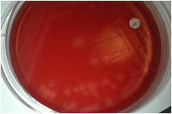
Numerous colony   morphologies ( hemolytic & non-hemolytic)

Non-hemolytic (γ-hemolysis): If an organism does not induce any hemolysis on a blood agar plate, it is said to display gamma or no hemolysis. The agar under and around the colony is unchanged.
Alpha hemolysis (α-hemolysis): Alpha hemolysis is the incomplete lysis of the red blood cells around and under the colonies on a blood agar plate. This area appears dark and greenish. Streptococcus pneumoniae and a group of streptococci (Streptococcus viridans or viridans streptococci) found in oral flora display alpha hemolysis.

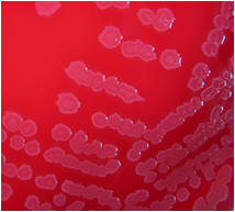
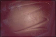
Beta hemolysis (β-hemolysis): Beta hemolysis is the complete lysis of the red blood cells around and under the colonies on a blood agar plate. This area appears transparent. Streptococcus pyogenes displays beta hemolysis and is often called Group A beta-hemolytic strep (GABHS).

Possible

Bacillus anthracis

 - Very fast growing

 - Non-hemolytic

 - Colony sticks together

 - Non-motile

Catalase test: The catalase test is used to differentiate some bacterial species. The test is done by placing a drop of hydrogen peroxide on a microscope slide. Using an applicator stick, a small portion of a colony is then added to a drop of hydrogen peroxide drop.

   Catalase +

Bacillus sp.

        Major pathogen: Bacillus anthracis

            Others only in immunocompromised patients from several blood cultures

@ Ellen Jo Baron 2007


Gram stain: The Gram stain, is a laboratory staining technique that distinguishes between two groups of bacteria that have differences in the structure of their cell walls. Standard bacterial taxonomy makes a distinction between Gram-negative bacteria, which stain red/pink and the Gram-positive bacteria, which stain blue/purple. Different antimicrobial agents are directed specifically at gram-positive bacteria and gram-negative bacteria.

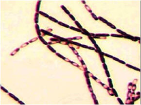
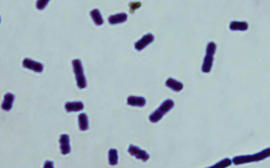
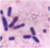
 Regular Gram + rods & spores (do not stain)

 B. anthracis

 from broth – central spores